Visual Processing and Visual Snow Syndrome
Visual System Anatomy
The Eye: Our Window to the World
Vision begins the moment light enters the eye. Think of the eye as a sophisticated camera, gathering light from the environment and starting the process of turning it into a picture you can understand. The first part of this journey happens at the eye's surface.
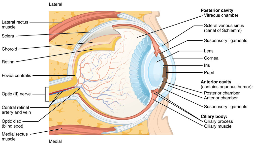
Light first passes through the cornea, a transparent, dome-shaped layer that covers the front of the eye. It acts like a protective window and does most of the light focusing.
Behind the cornea, light travels through the pupil and hits the lens. The lens is flexible and can change its shape to fine-tune the focus, allowing us to see objects clearly whether they are near or far. It directs the focused light toward the back of the eye.
The cornea provides most of the eye's focusing power, while the lens makes adjustments for distance.
The back of the eye is lined with a light-sensitive layer of tissue called the retina. This is where the magic happens. The retina contains millions of specialized photoreceptor cells, known as rods and cones, which convert light energy into electrical signals. It's the film in the camera, capturing the focused image.
retina
noun
A thin layer of tissue at the back of the eye that contains photoreceptor cells, which convert light into neural signals.
Once the retina captures the light and turns it into electrical signals, this information needs to get to the brain. This is the job of the optic nerve, a bundle of more than a million nerve fibers that acts as a high-speed data cable connecting the retina to the brain.
The Brain: Making Sense of Signals
The electrical signals travel along the optic nerve and arrive at a specific region in the back of the brain. The first stop in the cerebral cortex is an area called the primary visual cortex, often referred to as V1. It's located in the occipital lobe, right at the back of your head.
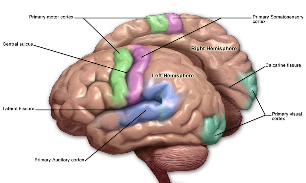
V1 acts as a sorting station. It begins to process the raw data from the eyes, detecting basic features like edges, lines, orientation, and movement. It doesn't see a whole object, like a face or a tree, but rather the simple components that make it up.
From V1, the visual information is sent to many other associated visual areas in the brain. These specialized regions work together to handle more complex tasks. Some areas are responsible for recognizing colors and shapes, while others help identify objects, recognize faces, or perceive motion and depth. It’s this coordinated effort that turns the simple lines and edges from V1 into the rich, detailed visual world we experience.
Now, let's test your knowledge of the visual system's anatomy.
Which part of the eye is a flexible structure that changes shape to fine-tune the focus of light?
The light-sensitive layer at the back of the eye, containing rods and cones, is called the __________.
Understanding this basic anatomy is the first step in exploring how we see.