Ventricular System and CSF Dynamics
Ventricular System Anatomy
The Brain's Inner Chambers
Deep within the brain lies a network of interconnected cavities known as the ventricular system. These spaces aren't empty; they are filled with cerebrospinal fluid (CSF), a clear liquid that cushions the brain, transports nutrients, and removes waste. Think of it as an internal hydraulic system for your central nervous system.
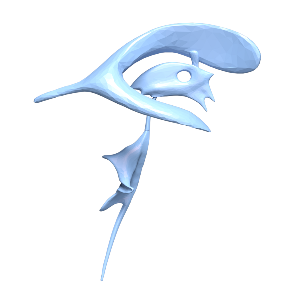
This system consists of four main chambers: two large lateral ventricles, a single third ventricle, and a single fourth ventricle. Each has a unique shape and location, and they are all linked by narrow passageways, allowing CSF to flow throughout the brain and spinal cord.
The Lateral Ventricles
The two lateral ventricles are the largest chambers, with one located in each cerebral hemisphere. They have a distinctive C-shape that curves from the frontal lobe, back through the parietal and occipital lobes, and then forward into the temporal lobe.
Because of this complex shape, each lateral ventricle is divided into three extensions, or "horns."
- Anterior horn: Projects into the frontal lobe.
- Posterior horn: Extends back into the occipital lobe.
- Inferior horn: Curves down and forward into the temporal lobe.
The two lateral ventricles connect to the third ventricle via a pair of small channels. Each channel is called an , also known by the eponym Foramen of Monro.
The Midline Ventricles
Situated in the middle of the brain, the third and fourth ventricles act as the central conduits of the system. The third ventricle is a narrow, slit-like cavity located in the region, nestled between the left and right thalamus. It receives CSF from both lateral ventricles through the foramina of Monro.
From the third ventricle, CSF flows downward through a long, narrow tube called the (or Aqueduct of Sylvius). This canal passes through the midbrain and connects the third ventricle to the fourth ventricle.
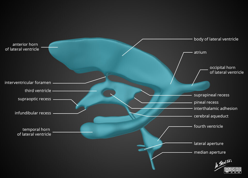
The fourth ventricle is a diamond-shaped chamber located posterior to the pons and upper medulla oblongata, and anterior to the cerebellum. Its position is critical, as it serves as the final gateway for CSF to exit the ventricular system and enter the subarachnoid space, which surrounds the entire brain and spinal cord.
This exit is made possible by three small openings, or apertures:
- Two lateral apertures (Foramina of Luschka): Located on the side walls of the ventricle.
- One median aperture (Foramen of Magendie): Found in the roof of the ventricle.
From the 4th ventricle, the fluid drains into two places:
Central spinal canal – bathes the spinal cord Subarachnoid cisterns – bathes the brain, between arachnoid mater and pia mater. Here the CSF is reabsorbed back into the circulation.
Once CSF passes through these apertures, it circulates around the brain and spinal cord before eventually being reabsorbed into the bloodstream. This constant flow ensures the central nervous system remains protected and chemically stable.
What is the primary function of the cerebrospinal fluid (CSF) that fills the ventricular system?
Which part of the lateral ventricle extends down and forward into the temporal lobe?
Understanding the brain's ventricular system provides a map for the flow of cerebrospinal fluid, a process vital to neurological health.