Ventricular Safety Pacing Explained
Cardiac Electrophysiology
The Heart’s Electrical Symphony
The heart is more than a muscular pump. It's a precise electrical organ. Every heartbeat is driven by a tiny, coordinated electrical impulse that sweeps through the heart muscle in a specific pattern. This ensures the chambers contract in the right sequence, efficiently pumping blood to the lungs and the rest of the body.
This entire process is managed by a dedicated network of specialized cells known as the cardiac conduction system. Think of it as the heart's internal wiring, responsible for creating and distributing the signals that keep it beating.
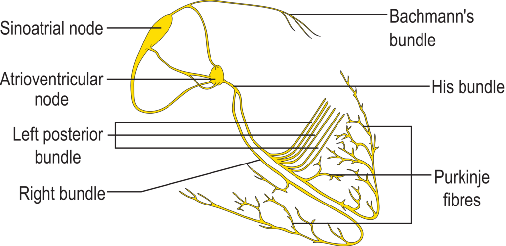
Let's trace the path of this electrical signal, starting from its source.
The Natural Pacemaker
The journey begins in the sinoatrial (SA) node, a small cluster of cells located in the upper wall of the right atrium. The SA node is the heart's natural pacemaker. It has a special ability to generate electrical impulses automatically, without any signal from the brain. In a resting adult, it typically fires about 60 to 100 times per minute, setting the pace for the entire heart.
Once the SA node fires, the electrical wave spreads out across both atria, like ripples in a pond. This wave of depolarization causes the atrial muscles to contract, pushing blood down into the ventricles.
The Conduction Pathway
The signal can't just jump directly from the atria to the ventricles. There's a layer of non-conductive tissue separating them. Instead, the signal funnels into the atrioventricular (AV) node, located near the center of the heart.
The AV node acts as a critical gatekeeper. It receives the signal from the atria and holds it for a fraction of a second. This slight delay is crucial; it gives the ventricles enough time to fill completely with blood from the contracting atria before they are told to pump.
After this brief pause, the AV node releases the signal down a pathway called the bundle of His. This bundle quickly splits into two branches, a right bundle branch and a left bundle branch, which run down along the septum, the muscular wall separating the right and left ventricles.
The AV node's delay ensures that the atria contract and empty before the ventricles contract and pump.
These bundle branches terminate in a vast network of tiny pathways called Purkinje fibers. These fibers spread throughout the muscular walls of the ventricles. They are designed for speed, rapidly distributing the electrical impulse to the entire ventricular muscle mass almost simultaneously. This triggers a powerful, coordinated contraction that starts at the bottom of the ventricles and moves upward, efficiently ejecting blood into the aorta and pulmonary artery.
When the Rhythm Fails
This entire sequence, from SA node to Purkinje fibers, happens with every single heartbeat, all in less than a quarter of a second. It's a remarkably reliable system, but it's not foolproof.
An arrhythmia is any disturbance in the rate, regularity, or pathway of this electrical impulse. If the SA node fires too fast or too slow, the heart rate changes. If the signal is blocked at the AV node, it can disrupt the coordination between the atria and ventricles. If an irritable group of cells somewhere else in the heart decides to fire on its own, it can create an extra, premature beat. These disruptions can range from harmless flutters to life-threatening emergencies, all stemming from a problem in the heart's electrical wiring.
Let's check your understanding of the heart's conduction system.
What is considered the natural pacemaker of the heart, responsible for initiating the electrical impulse for a normal heartbeat?
What is the primary purpose of the brief delay in signal transmission at the atrioventricular (AV) node?
A solid grasp of this electrical pathway is the first step toward understanding how the heart functions and what happens when things go wrong.