Venous System of the Lower Limb for Medical Exams
Superficial Venous Systems
Great Saphenous Vein
The Great Saphenous Vein (GSV) is the longest vein in the body. It originates from the dorsal venous arch of the foot, specifically from the union of the dorsal vein of the great toe and the arch itself. This vessel then ascends anterior to the medial malleolus, the bony prominence on the inner side of your ankle. This specific landmark is crucial for locating the vein for clinical procedures like cannulation.
From the ankle, the GSV continues its course up the medial side of the leg, just posterior to the medial border of the tibia. As it crosses the knee, it runs posterior to the medial condyle of the femur. It then continues superiorly along the medial aspect of the thigh. Throughout its journey, it contains between 10 and 20 valves, which are more numerous in the leg section than in the thigh, to prevent the backflow of blood.
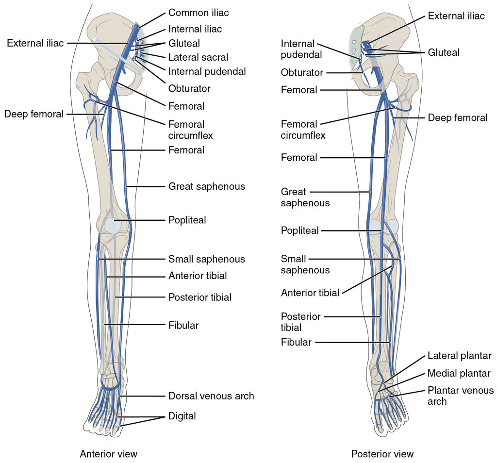
The GSV's journey concludes as it dives deep to join the femoral vein. It does this by passing through a specific aperture in the fascia lata of the thigh called the , also known as the fossa ovalis. This meeting point, the , is located about 3-4 centimeters inferolateral to the pubic tubercle. Just before its termination, the GSV receives several key tributaries: the superficial circumflex iliac vein, the superficial epigastric vein, and the external pudendal veins. An anterolateral accessory saphenous vein and a posteromedial accessory saphenous vein also commonly drain into it within the thigh.
Small Saphenous Vein
The Small Saphenous Vein (SSV), also known as the lesser saphenous vein, originates on the other side of the foot. It's formed by the union of the dorsal vein of the little toe with the dorsal venous arch. From its origin, it travels posterior to the lateral malleolus, accompanying the sural nerve.
The SSV then ascends along the posterior aspect of the leg, positioned between the two heads of the gastrocnemius muscle. It contains approximately 7 to 13 valves to aid in unidirectional blood flow. Its termination point is variable, but it most commonly pierces the deep popliteal fascia and drains into the within the popliteal fossa, the diamond-shaped space behind the knee.
Anatomical variation is common. In some individuals, the SSV may join the GSV, or it might continue up the thigh as the vein of Giacomini to drain into the femoral vein.
Valves and Flow
The valves in both the GSV and SSV are bicuspid, consisting of two delicate leaflets that coapt to prevent retrograde blood flow. The highest concentration of valves is found in the distal portions of the veins (closer to the ankle), where hydrostatic pressure is greatest. The function of these superficial veins is critically supported by the calf muscle pump. As the gastrocnemius and soleus muscles contract during walking, they squeeze the deep veins of the leg, propelling blood upward. This action reduces pressure in the deep system, creating a gradient that encourages blood to flow from the superficial system (GSV and SSV) into the deep system through perforator veins.
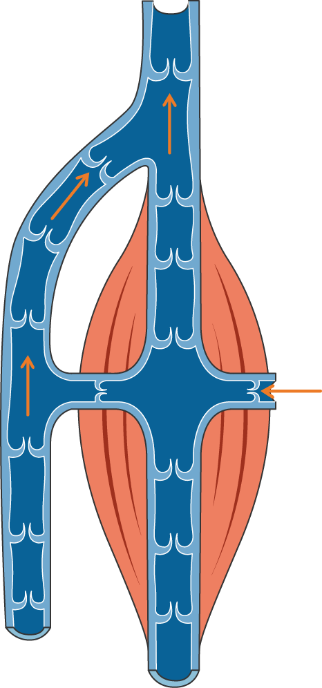
When these valves fail, blood pools in the veins, leading to increased pressure and venous hypertension. This can result in conditions like varicose veins, where the vessels become swollen, twisted, and visible beneath the skin.
Where does the Great Saphenous Vein (GSV) originate?
To locate the Great Saphenous Vein for a clinical procedure near the ankle, which bony landmark is the most accurate reference?
You've now covered the detailed anatomy of the major superficial veins of the lower limb. These pathways are essential for both normal physiological function and a variety of clinical scenarios.