Understanding the TMJ
TMJ Anatomy
Your Jaw's Hinge
Every time you talk, chew, or yawn, you're using your temporomandibular joints, or TMJs. You have two of them, one on each side of your head, acting as sliding hinges connecting your jawbone to your skull. They are among the most complex joints in the body, capable of a wide range of motion. To understand how they work, let's break them down into their core components: bones, muscles, ligaments, and a special little disc.
The Bony Framework
The TMJ is where two specific bones meet. The first is the lower jaw, known as the mandible. The part of the mandible that forms the joint is a rounded knob called the mandibular condyle.
The condyle fits into a shallow socket on the skull. This socket is part of the temporal bone (the same bone where your ear is located) and is called the mandibular fossa. These two bony structures, the condyle and the fossa, are the foundation of the joint.
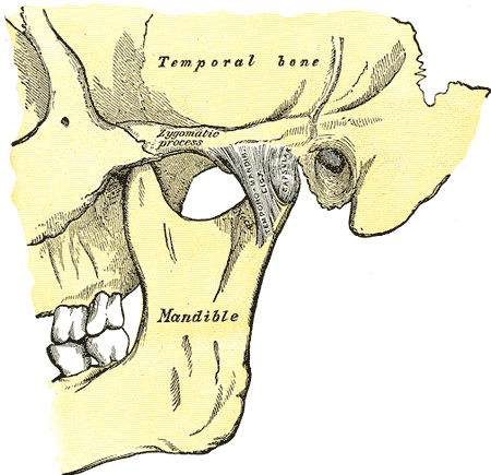
The articular disc is a small, tough, and flexible piece of fibrocartilage that sits right between these two bones. Think of it as a tiny, movable cushion. It's not attached to bone directly but is held in place by ligaments.
This disc is crucial. It absorbs shock from chewing and allows the jaw to move smoothly. Without it, the bones would grind against each other.
The disc also divides the joint into two separate compartments or synovial cavities, an upper and a lower one. Each compartment is filled with synovial fluid, a natural lubricant. This two-part structure allows the TMJ to perform its unique combination of hinging and gliding movements. The lower compartment handles the initial hinging motion when you first open your mouth, while the upper compartment allows the jaw to glide forward and down.
Muscles and Ligaments
Bones don't move on their own. The TMJ is controlled by a powerful group of muscles known as the muscles of mastication (chewing). These muscles work in pairs to move the mandible in different directions.
| Muscle | Primary Action |
|---|---|
| Masseter | Closes the jaw (elevation) |
| Temporalis | Closes the jaw (elevation) and pulls it back (retraction) |
| Medial Pterygoid | Closes the jaw (elevation) and helps with side-to-side movement |
| Lateral Pterygoid | Opens the jaw (depression) and pushes it forward (protrusion) |
These muscles provide the force for movement, but ligaments provide stability. Ligaments are tough, fibrous bands that connect bones and prevent the joint from moving too far. The TMJ has several key ligaments that enclose the joint and limit its range of motion, ensuring the condyle stays within the fossa during movement.
Together, these components—bones, disc, muscles, and ligaments—work in harmony to allow for the complex movements required for everyday life. Understanding this intricate anatomy is the first step in appreciating how this small but mighty joint functions.