Understanding Peptic Ulcer Disease Pathology
Gastrointestinal Tract Anatomy
The Upper GI Tract
The upper gastrointestinal (GI) tract is where digestion truly kicks into high gear. After you chew and swallow, food travels down the esophagus and enters the stomach, a muscular, J-shaped organ tucked in the upper abdomen. The stomach's main job is to act like a blender, churning food with powerful acids and enzymes to break it down into a semi-liquid mixture called chyme.
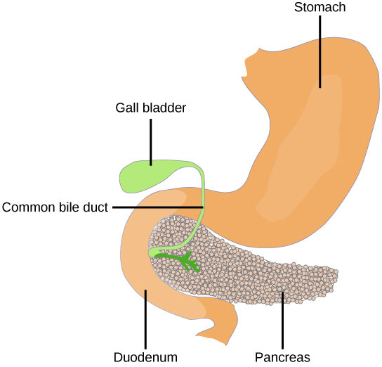
The stomach has several distinct regions. The top, rounded part is the fundus. The main, central section is the body. The lower, funnel-shaped portion is the antrum, which connects to the small intestine through a muscular valve called the pyloric sphincter. This sphincter controls how much chyme is released and when.
Inside, the stomach lining isn't smooth. It's covered in deep folds.
Most of the gastric mucosa is thrown up in coarse folds called rugae, while the antral mucosa is much smoother.
These rugae allow the stomach to expand significantly to hold a large meal.
The Duodenum
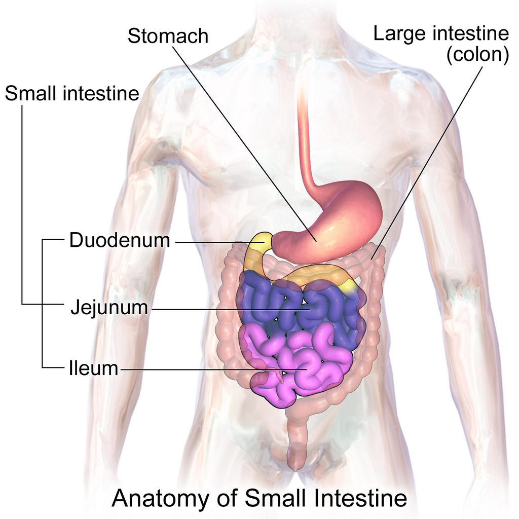
Once the chyme is ready, it passes into the duodenum, the first and shortest section of the small intestine. It's a C-shaped tube that curves around the head of the pancreas. The duodenum is a critical mixing bowl where chyme from the stomach is combined with bile from the gallbladder and digestive enzymes from the pancreas. This process neutralizes stomach acid and continues the breakdown of proteins, fats, and carbohydrates.
A Four-Layer Wall
The entire GI tract, from the stomach onward, shares a similar four-layered structure. Think of it as a tube made of concentric rings, each with a specific function.
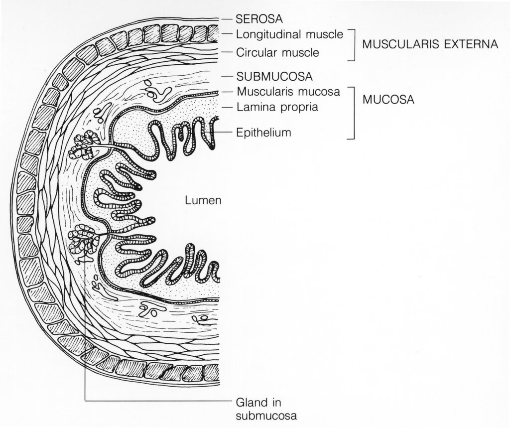
From the inside out, the layers are:
- Mucosa: This is the innermost layer that comes into direct contact with food. It's responsible for secretion and absorption. The mucosa itself has three sublayers: an epithelial lining, a connective tissue layer called the lamina propria, and a thin muscle layer called the muscularis mucosae.
- Submucosa: This layer contains blood vessels, lymphatic vessels, nerves, and glands that support the mucosa.
- Muscularis Externa: This is the main muscle layer responsible for peristalsis, the wave-like contractions that move food through the GI tract. It typically has an inner circular layer and an outer longitudinal layer.
- Serosa: The outermost layer is a thin, slippery membrane that lubricates the outside of the GI tract, allowing organs to slide past each other without friction.
The Mucosal Shield
The environment inside the stomach is incredibly harsh. Gastric acid has a pH between 1.5 and 3.5, which is strong enough to dissolve metal. So how does the stomach avoid digesting itself? It relies on a sophisticated protective barrier.
The first line of defense is a thick layer of mucus secreted by specialized cells in the epithelial lining. This mucus coats the stomach wall, creating a physical barrier between the acidic chyme and the delicate cells underneath.
But mucus alone isn't enough. Embedded within this mucus layer is a chemical shield.
Epithelial cells also secrete bicarbonate, an alkaline substance that neutralizes any acid that manages to seep through the mucus. This creates a pH-neutral zone right at the surface of the cells, keeping them safe. This dynamic duo of mucus and bicarbonate is essential for maintaining the integrity of the gastric and duodenal lining.
What is the primary function of the stomach in digestion?
What is the correct order of the four main layers of the GI tract, from the innermost layer to the outermost layer?
Understanding this basic anatomy is the first step. Next, we'll explore what happens when these protective mechanisms are compromised.