Understanding Heart Sounds
Heart Anatomy
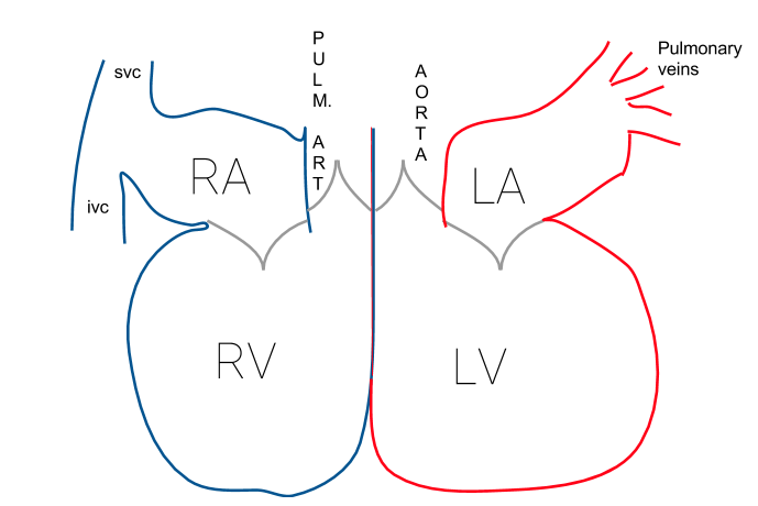
The Heart's Four Chambers
The heart is a powerful muscle, working tirelessly to pump blood throughout your body. It's organized into four distinct rooms, or chambers, each with a specific job. Think of it as a two-story duplex, with a right side and a left side.
The two upper chambers are called the atria (singular: atrium). They act as receiving rooms. The right atrium collects oxygen-poor blood returning from the body. The left atrium collects oxygen-rich blood coming from the lungs.
The two lower chambers are the ventricles. These are the powerful pumps. The right ventricle pumps the oxygen-poor blood it receives from the right atrium to the lungs. The left ventricle is the strongest chamber; it pumps the oxygen-rich blood it gets from the left atrium out to the rest of the body.
One-Way Doors: The Heart Valves
To keep blood flowing in the right direction, the heart has four valves. These valves act like one-way doors, opening to let blood pass through and then snapping shut to prevent it from flowing backward.
There are two main types:
-
Atrioventricular (AV) valves: These sit between the atria and the ventricles.
- The tricuspid valve is on the right side, between the right atrium and right ventricle.
- The mitral valve is on the left side, between the left atrium and left ventricle.
-
Semilunar valves: These are at the exits of the ventricles.
- The pulmonary valve is between the right ventricle and the pulmonary artery.
- The aortic valve is between the left ventricle and the aorta.
The Circulatory Highway
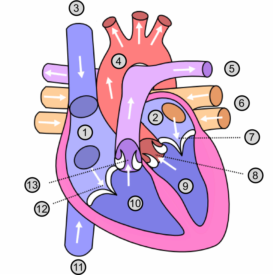
A network of major blood vessels connects to the heart, forming the main highways of your circulatory system. These vessels are responsible for carrying blood to and from the heart.
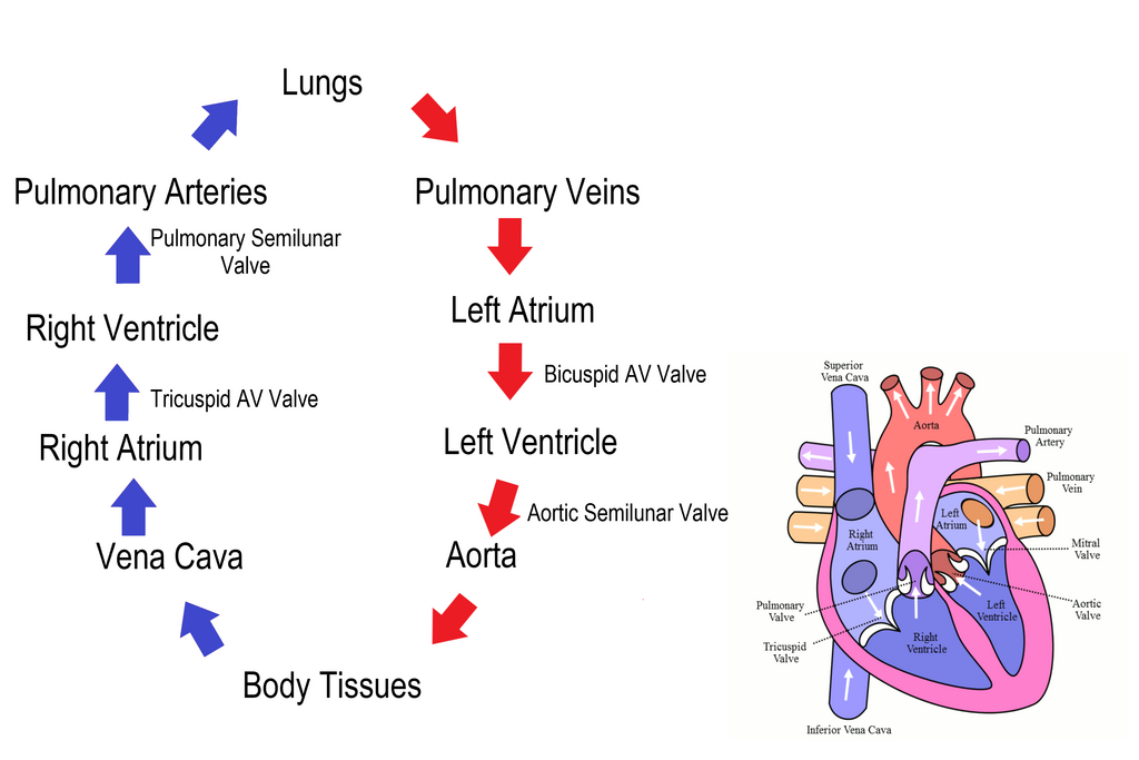
Deoxygenated blood (blue) comes from the body, goes to the lungs, and returns as oxygenated blood (red) to be pumped back out to the body.
Here are the key players:
- The superior and inferior vena cava are two large veins that bring oxygen-poor blood from the body into the right atrium.
- The pulmonary artery carries that oxygen-poor blood from the right ventricle to the lungs to pick up oxygen.
- The pulmonary veins carry oxygen-rich blood from the lungs back to the left atrium.
- The aorta is the body's largest artery. It carries oxygen-rich blood from the left ventricle to the rest of the body.
The Rhythm of the Pump
The continuous sequence of contraction and relaxation of the heart is called the cardiac cycle. This cycle is divided into two main phases, which create the familiar "lub-dub" sound of a heartbeat.
Systole
noun
The phase of the heartbeat when the heart muscle contracts and pumps blood from the chambers into the arteries.
Systole is the contraction phase. The ventricles squeeze, pushing blood out into the pulmonary artery and the aorta. This forceful contraction slams the mitral and tricuspid valves shut, creating the first heart sound (S1), the "lub."
Diastole
noun
The phase of the heartbeat when the heart muscle relaxes and allows the chambers to fill with blood.
Diastole is the relaxation phase. The ventricles relax and fill with blood from the atria. As they relax, the pressure change causes the aortic and pulmonary valves to snap shut. This creates the second heart sound (S2), the "dub."
This cycle of systole and diastole repeats with every beat, ensuring a continuous flow of blood.
Now that you know the parts of the heart and how they work together, you have the foundation for understanding heart sounds. Let's review the key terms.
Time to check your understanding of the heart's anatomy and function.
Which chamber of the heart is the most muscular, responsible for pumping oxygen-rich blood to the entire body?
The "lub" sound (S1) of a heartbeat is caused by the closing of which two valves?
Understanding this anatomy is the first step in learning to interpret the sounds the heart makes. Each chamber, valve, and vessel plays a role in the symphony of the cardiac cycle.