Ultrasonography for Pregnancy Diagnosis in Sheep and Goats
Restraint and Scanning Workflows
Setting the Stage for Scanning
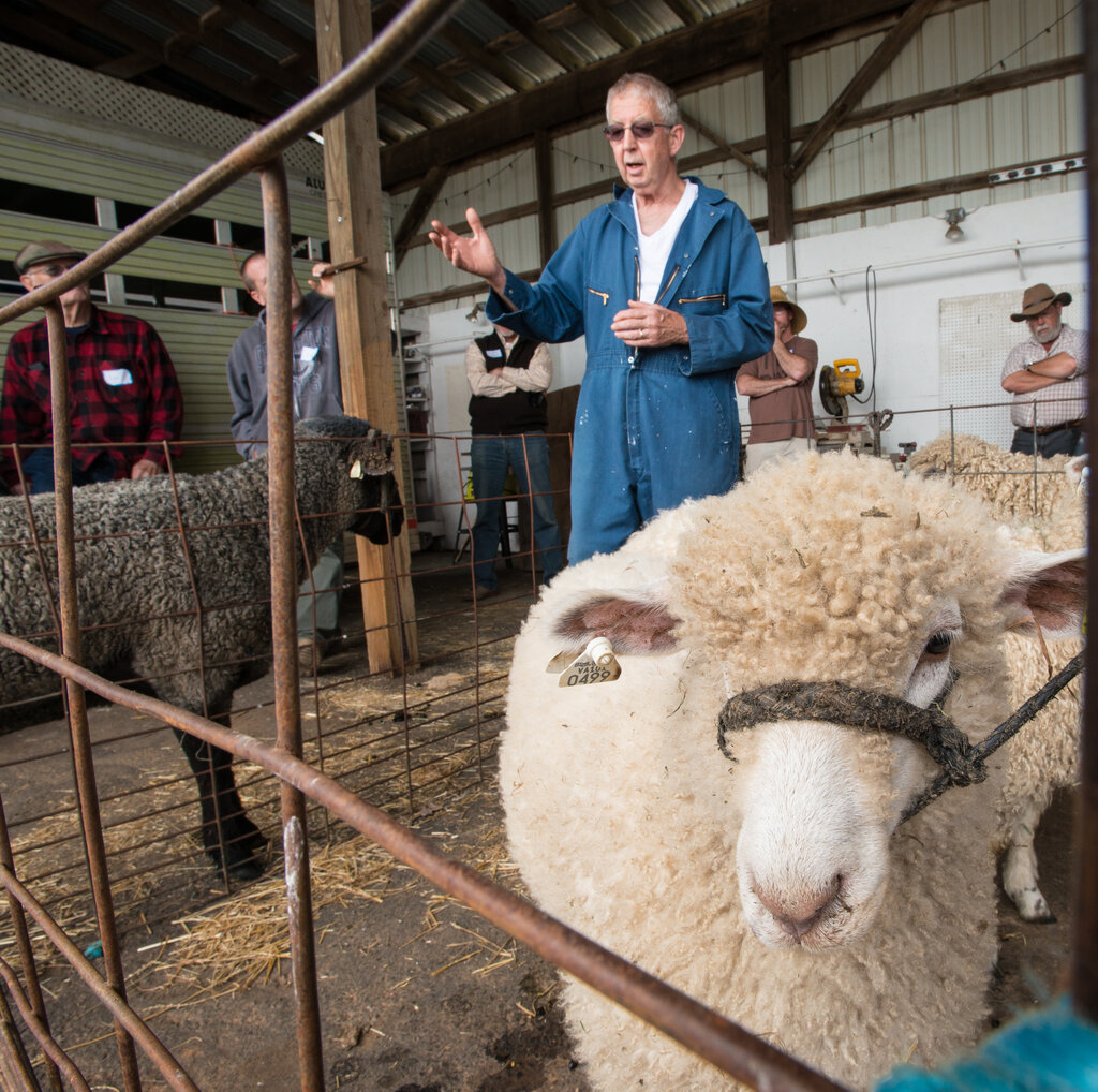
A successful ultrasound exam begins long before the probe touches the animal. Proper restraint is non-negotiable. A stressed, moving animal leads to a frustrated operator and poor-quality, unreliable images. The goal is to secure the ewe or doe comfortably and safely, limiting movement without causing panic.
For small-scale operations, manual restraint can be effective. One person can hold the animal steady against a wall or in a corner. For larger flocks, dedicated equipment is a wise investment. A milking stand, stanchion, or specialized head gate can immobilize the animal's head and body, leaving the operator free to focus on the scan. Some producers use a 'deck chair' or 'tipping table' that gently lays the animal on its back, providing excellent access to the abdomen.
Choosing Your Approach
With the animal secured, you have two primary methods for pregnancy detection, each suited to a different stage of gestation.
The transrectal approach is the go-to method for early confirmation, typically between days 18 and 40. This technique involves inserting a high-frequency (5-7.5 MHz) linear probe into the rectum. Due to the small working area and potential risk to the operator, using probe extenders is highly recommended. These devices add length and rigidity, allowing for safer and more controlled manipulation of the probe to visualize the uterus.
For mid-to-late gestation (day 40 onwards), the transabdominal approach is standard. As the fetus grows and the uterus expands, it drops down into the abdominal cavity, making it easily visible from the outside. This method uses a lower-frequency (3.5-5 MHz) sector or convex probe, which provides a wider field of view.
A critical detail for this technique is to scan from the animal's right side. The left side is dominated by the a large digestive organ that will block the ultrasound waves and obscure your view of the uterus. By scanning from the right, in the hair-free inguinal region just in front of the udder, you get a clear acoustic window to the reproductive tract.
Getting a Clear Picture
Ultrasound waves travel poorly through air. To get a clear image, you need a coupling agent to ensure solid contact between the probe and the animal's skin. For transabdominal scanning, liberally apply either isopropyl alcohol or acoustic gel to the hair-free inguinal area. Alcohol is inexpensive and works well, but it can evaporate quickly. Gel provides longer-lasting contact but can be messy and collect dirt.
For transrectal exams, a generous amount of obstetric lubricant is essential for both image quality and the animal's comfort.
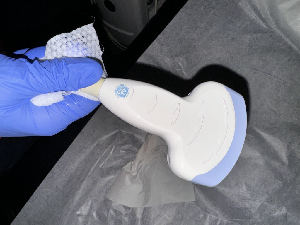
Once you have contact, you'll need to fine-tune the machine's settings. While every machine is different, focus on three main controls:
- Frequency: As discussed, use higher frequencies (5-7.5 MHz) for detailed, shallow structures (transrectal) and lower frequencies (3.5-5 MHz) for deeper penetration (transabdominal).
- Depth: Adjust this so your area of interest fills about 75% of the screen. Too deep, and the image is too small. Too shallow, and you might miss what you're looking for.
- : This controls the overall brightness of the image. Adjust the gain until you have a clear image with good contrast between tissues and fluid. Fluid-filled structures, like an early pregnancy vesicle or the fetal bladder, should appear black (anechoic).
A systematic approach is key. Always start with the same restraint method, prepare the site thoroughly, and follow a consistent pattern as you move the probe to examine the anatomy.
Ready to test your knowledge on setting up for a scan?
What is the primary reason for properly restraining a ewe or doe before an ultrasound exam?
When performing a transabdominal ultrasound on a pregnant ewe, why is it critical to scan from the animal's right side?
Mastering these physical workflows is the foundation for everything that follows. With the animal properly restrained and your machine optimized, you're ready to interpret the images on the screen.