TL1A and the AI Revolution in IBD Target Discovery
TL1A DR3 Signaling Mechanics
The TL1A-DR3 Signaling Axis
In the complex network of immune communication, some signals act like a quiet conversation, while others are a shout. The interaction between (TL1A) and its receptor, Death Receptor 3 (DR3), is definitely a shout. TL1A, also known as TNFSF15, is a protein expressed by various immune cells. It has only one known partner: DR3. This exclusivity makes their connection, or signaling axis, a highly specific and potent driver of immune responses.
Think of it as a lock and key. DR3 is the lock, found on the surface of specific lymphocytes, particularly activated T cells. TL1A is the key, presented by antigen-presenting cells (APCs) like macrophages and dendritic cells. When the key turns the lock, it doesn't just open a door; it kicks off a powerful cascade that can escalate an immune response from a controlled skirmish into chronic inflammation. This is especially relevant in inflammatory bowel disease (IBD), where this axis is often in overdrive.
A Potent Boost for T Cells
T-cell activation isn't a simple on-off switch. For a CD4+ T cell to become fully activated, it needs two distinct signals. The first comes from its T-cell receptor (TCR) recognizing a specific antigen presented by an APC. But this signal alone is often weak, leading to an ineffective response or even T-cell anergy (a state of shutdown).
To get a robust response, a second, co-stimulatory signal is required. This is where TL1A comes in. When an APC presents an antigen to a T cell, it can also present TL1A on its surface. The TL1A binds to the DR3 receptor on the T cell, providing a powerful co-stimulatory jolt. This secondary signal dramatically enhances the initial TCR signal, pushing the T cell to proliferate rapidly and differentiate into an effector cell ready to fight.
Activating Inflammatory Pathways
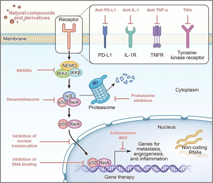
Once TL1A binds to DR3, a series of events is triggered inside the T cell. The DR3 receptor recruits adapter proteins, initiating a domino effect that activates two key intracellular signaling pathways: NF-κB and MAPK. These aren't just random acronyms; they are master regulators of gene expression for inflammation. The , in particular, is a central hub for immune cell activation and survival.
Activation of NF-κB and MAPK pathways effectively tells the T cell's nucleus to start producing and secreting pro-inflammatory cytokines. The two most notable cytokines produced under TL1A's influence are Interferon-gamma (IFN-γ) and Tumor Necrosis Factor-alpha (TNF-α). These molecules are potent drivers of the inflammation that damages tissue in the gut of IBD patients.
The Power of Synergy
TL1A signaling is powerful on its own, but its inflammatory potential is truly unlocked when it works in concert with other signals. In the gut, T cells are often exposed to a cocktail of cytokines. Two of these, (IL-12) and Interleukin-23 (IL-23), have a powerful synergistic relationship with TL1A.
When a T cell receives a signal from TL1A at the same time it's being stimulated by IL-12 or IL-23, the outcome is more than just additive. The combined signals create an amplified inflammatory response, strongly promoting the expansion of highly pathogenic T-cell subsets like Th1 and Th17 cells. This synergy is a key reason why the TL1A-DR3 axis is so central to the chronic, self-sustaining inflammation seen in IBD. It turns up the volume on other inflammatory signals, creating a vicious cycle of tissue damage.
By understanding these specific molecular mechanics, from the initial APC interaction to the activation of intracellular pathways and cytokine synergy, we can see why targeting the TL1A-DR3 axis is a promising strategy for treating IBD.