Tissue Harmonics in Ultrasound
Ultrasound Physics
The Sound You Can't Hear
Sound is simply a vibration traveling through a medium, like air or water. We perceive these vibrations as sound when they reach our ears. Ultrasound is the same kind of wave, but its frequency is too high for humans to hear. While we can typically hear sounds up to about 20,000 Hertz (Hz), medical ultrasound uses frequencies in the range of 2 to 15 million Hertz (MHz).
Think of it as a dog whistle. A dog can hear it, but for you, it's just silence. Ultrasound works on a similar principle, using high-frequency sound waves to peek inside the body without any cutting or radiation.
But how do you create a sound that high-pitched? The magic lies in special materials with a unique ability.
Crystals That Create Sound
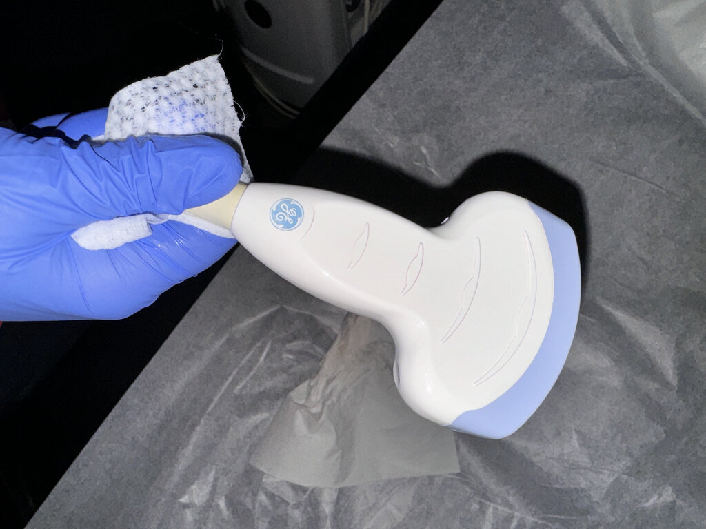
The heart of an ultrasound machine is the transducer, a small handheld device placed on the skin. Inside it are tiny piezoelectric crystals. The term comes from the Greek word piezein, which means “to press.”
These crystals have a remarkable property: when you apply an electric voltage to them, they change shape. By applying a rapidly alternating voltage, the crystals vibrate back and forth very quickly, creating pressure waves—the ultrasound waves. These waves then travel from the transducer into the body.
The process also works in reverse. When returning sound waves (echoes) hit the crystals, they generate an electric voltage. This signal is sent back to the computer to form an image.
Wave Properties
Once created, these waves travel into the body. To understand how they build an image, we need to know a few of their basic properties.
Frequency
noun
The number of wave cycles that pass a point per second. Measured in Hertz (Hz).
Wavelength
noun
The physical distance over which one complete wave cycle occurs.
Frequency (), wavelength (), and the speed of the wave () are all related. The speed of sound is determined by the medium it travels through. In the body's soft tissues, it's remarkably consistent, averaging about 1540 meters per second.
Since the speed of sound in tissue is relatively fixed, this formula tells us that frequency and wavelength are inversely proportional. A high-frequency wave has a short wavelength, and a low-frequency wave has a long wavelength. This trade-off is fundamental to ultrasound imaging.
Interactions Inside the Body
An ultrasound image is essentially a map of how sound waves interact with different tissues. These interactions are governed by a property called acoustic impedance.
Acoustic Impedance
noun
A physical property of a medium that describes its resistance to the passage of sound waves.
Acoustic impedance () is calculated by multiplying the density of the tissue () by the speed of sound in that tissue ().
Every type of tissue—fat, muscle, liver, bone—has a different acoustic impedance. When an ultrasound wave traveling through one tissue hits the boundary of another, this difference in impedance causes some of the wave to bounce back, while the rest continues deeper. These returning echoes are what create the image.
| Interaction | Description |
|---|---|
| Reflection | The wave bounces back from a boundary between tissues. This creates the image. A large impedance difference (like between muscle and bone) creates a strong, bright echo. |
| Refraction | The wave bends as it passes from one tissue to another. This is similar to how light bends in water. |
| Attenuation | The wave loses energy and becomes weaker as it travels deeper into the body. This is why it's harder to image structures that are very deep. |
By timing how long it takes for the echoes to return and measuring their strength, the ultrasound machine can calculate the depth and properties of different structures, piecing them together to form a detailed, real-time image.
What fundamental property distinguishes ultrasound from the sound humans can normally hear?
The generation of ultrasound waves in a medical transducer relies on which physical phenomenon?