Tibia Anatomy and Biomechanics
Structural Functional Anatomy
The Tibia's Architecture
The tibia, or shin bone, is the primary weight-bearing bone of the lower leg. It acts as a crucial pillar, transferring the body's weight from the femur above to the talus bone in the ankle below. Its design, however, is far more sophisticated than that of a simple post. The tibia's internal and external architecture is a masterclass in balancing strength, weight, and the ability to grow and repair itself.
The lower limb supports body weight, provides stability and enables locomotion.
To understand how it handles immense daily stress, we need to look at its material composition. The tibia is built from two main types of bone tissue: dense cortical bone and spongy cancellous bone. The distribution of these materials is not random; it's strategically organised to maximise support where it's needed most.
The long central shaft, or diaphysis, is composed of a thick tube of cortical bone. This provides the rigidity required to resist bending and twisting forces. At the proximal and distal ends (the epiphyses), the structure changes. Here, a thinner shell of cortical bone surrounds a core of cancellous bone. This honeycomb-like structure is lighter but excellent at absorbing and distributing the compressive forces that occur at the knee and ankle joints.
An Engineered Cross-Section
If you were to slice through the tibial shaft, you wouldn't see a perfect circle. Instead, it has a distinct triangular cross-section. This shape is a brilliant piece of natural engineering. A triangular profile provides superior resistance to bending forces from multiple directions compared to a circular shape of the same mass. It offers strength against forces from the front, back, and sides, all while minimising bone weight.
This structural efficiency is vital. It allows the tibia to be strong enough for running and jumping without being excessively heavy, which would require more energy to move. The interior of this cortical shaft isn't solid, either. It contains the medullary canal.
The medullary canal is a hollow cavity that runs the length of the diaphysis. It's filled with bone marrow and plays a role in distributing pressure within the bone. By hollowing out the centre, the bone's weight is significantly reduced with minimal loss of bending strength, another principle often seen in mechanical engineering.
Lifeblood and Growth
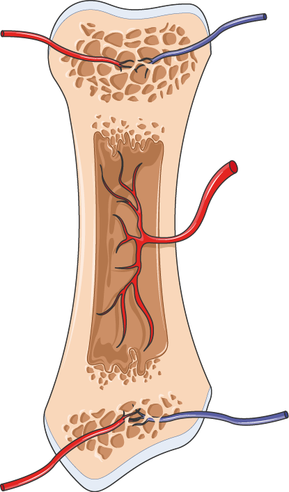
Like any living tissue, bone requires a constant blood supply to deliver nutrients and remove waste. The tibia's primary vessel is the nutrient artery, which enters the bone through a small opening called the nutrient foramen. This foramen is typically located on the posterior surface of the upper third of the tibia.
Its location and integrity are clinically significant. In the event of a fracture, a preserved blood supply via the nutrient artery is critical for successful healing. Surgeons must be mindful of this vessel's location during procedures to avoid disrupting the bone's primary source of nourishment.
A bone's architecture is not static from birth. Long bones like the tibia grow in length from specific areas near their ends called epiphyseal plates, or growth plates. These are regions of cartilage where ossification, the process of bone formation, occurs.
The tibia has two main ossification centres: a primary centre in the diaphysis that appears before birth, and secondary centres that appear in the proximal and distal epiphyses after birth. The growth plates remain active throughout childhood and adolescence, adding length to the bone. Eventually, in early adulthood, these plates close and are replaced by solid bone, marking the end of longitudinal growth.
What is the primary function of the tibia?
The triangular cross-section of the tibial shaft is advantageous because it...