The Sliding Filament Theory of Muscle Contraction
Muscle Structure
The Architecture of Muscle
Your muscles are complex structures, organized in a series of bundles within bundles. Think of a large rope made of smaller ropes, which are in turn made of even finer threads. A whole muscle is wrapped in a protective layer of connective tissue called the epimysium. Inside, the muscle is sectioned into bundles called fascicles, each wrapped in another layer called the perimysium.
Drill down further and you'll find the individual muscle fibers within each fascicle. These aren't just threads; they are the actual muscle cells. Each fiber is a single, long cell that can be quite large, and unusually, contains multiple nuclei. A delicate layer of connective tissue, the endomysium, surrounds each individual muscle fiber, providing support and pathways for nerves and blood vessels.
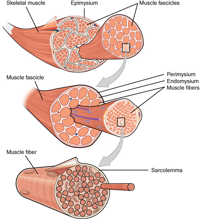
Inside each muscle fiber are even smaller structures called myofibrils. These are long, cylindrical organelles that run the entire length of the muscle fiber. The myofibrils are the real workhorses of the muscle cell, and their unique structure is what allows muscles to contract.
The Sarcomere
If you could zoom in on a single myofibril, you would see a repeating pattern of light and dark bands. This striated, or striped, appearance is a hallmark of skeletal muscle. Each repeating unit of this pattern is called a sarcomere.
The sarcomere is the fundamental contractile unit of a muscle fiber. A single myofibril is essentially a long chain of sarcomeres linked end to end.
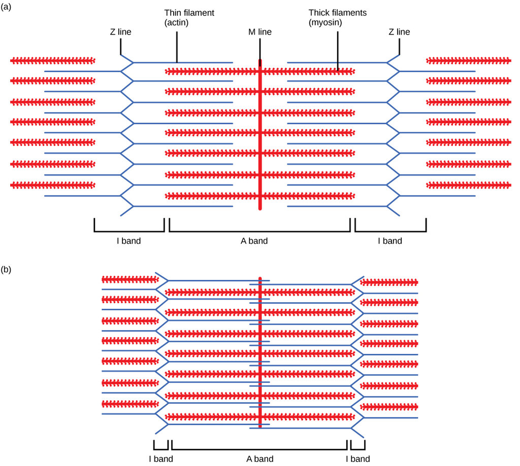
The boundaries of a sarcomere are marked by structures called Z-lines (or Z-discs). Running from one Z-line to the next, you can observe several distinct regions:
- I-Band: A light-colored band located on either side of the Z-line. It contains only thin filaments.
- A-Band: A dark, central region that spans the entire length of the thick filaments. The outer edges of the A-band are where thick and thin filaments overlap.
- H-Zone: A slightly lighter area within the middle of the A-band. This zone contains only thick filaments.
- M-Line: A line found in the very center of the H-zone, which helps hold the thick filaments in place.
Thick and Thin Filaments
The bands and lines of the sarcomere exist because of the orderly arrangement of two types of protein structures, called myofilaments.
Myosin
noun
The protein that makes up the thick filaments in muscle fibers. Myosin filaments have small 'heads' that can interact with actin.
The thick filaments are composed of the protein myosin. These filaments are anchored at the M-line and extend across the A-band. They have tiny projections, called myosin heads, which are the key to muscle movement.
Actin
noun
A protein that forms the thin filaments in muscle fibers. It provides binding sites for myosin heads during muscle contraction.
The thin filaments are primarily made of the protein actin. These filaments are attached to the Z-lines and extend inward across the I-band and partway into the A-band, where they overlap with the thick filaments. It's the precise, overlapping arrangement of these actin and myosin filaments within millions of sarcomeres that gives a muscle its ability to produce force.