The Science of Breathing
Respiratory System Anatomy
The Upper Airway
Every breath you take begins a journey through a series of specialized passages. It all starts in the nasal cavity, the space behind your nose. This isn't just an empty tunnel; it's lined with a mucous membrane and tiny hairs called cilia. As you inhale, the air is warmed, humidified, and filtered, trapping dust and other particles before they can go any deeper.
From the nasal cavity, air moves into the pharynx, a muscular tube more commonly known as the throat. The pharynx is unique because it serves both the respiratory and digestive systems, acting as a pathway for air on its way to the lungs and for food on its way to the stomach. It's divided into three sections: the nasopharynx (behind the nose), the oropharynx (behind the mouth), and the laryngopharynx (the lowest part).
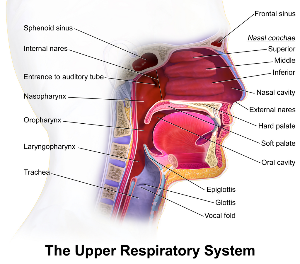
At the bottom of the pharynx, air enters the larynx, or voice box. The larynx is a short section of the airway made of cartilage. It houses the vocal cords, which vibrate to produce sound. Crucially, the larynx also has a gatekeeper: a flap of cartilage called the epiglottis. When you swallow, the epiglottis closes over the larynx to prevent food and drink from entering your windpipe.
The Lower Airway
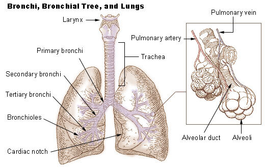
Past the larynx, air enters the lower respiratory tract, starting with the trachea, or windpipe. This is a sturdy tube, about 4-5 inches long, held open by C-shaped rings of cartilage. These rings provide support and prevent the trachea from collapsing.
The trachea travels down the chest and splits into two smaller tubes called the primary bronchi. There's one bronchus for each lung. This is where the airway begins to branch out like a tree.
Inside the lungs, the bronchi continue to divide, creating a vast network of smaller and smaller tubes. These are the secondary and tertiary bronchi, which lead to even narrower passages called bronchioles. Unlike the trachea and bronchi, bronchioles don't have cartilage rings. Their walls are made of smooth muscle, which allows their diameter to change.
The Lungs and Alveoli
The branching network of bronchioles finally terminates in the functional units of the lungs: the alveoli. The lungs themselves are two spongy, cone-shaped organs located in the chest cavity. The right lung is slightly larger and has three lobes, while the left lung has two lobes to make room for the heart.
At the very end of the tiniest bronchioles are clusters of microscopic air sacs that look like bunches of grapes. These are the alveoli. There are hundreds of millions of them in your lungs. Their walls are incredibly thin, just a single cell thick, and they are surrounded by a dense network of tiny blood vessels called capillaries. This intimate contact between air and blood is the key to respiration, though the process of gas exchange itself is a story for another time.
Protective Linings
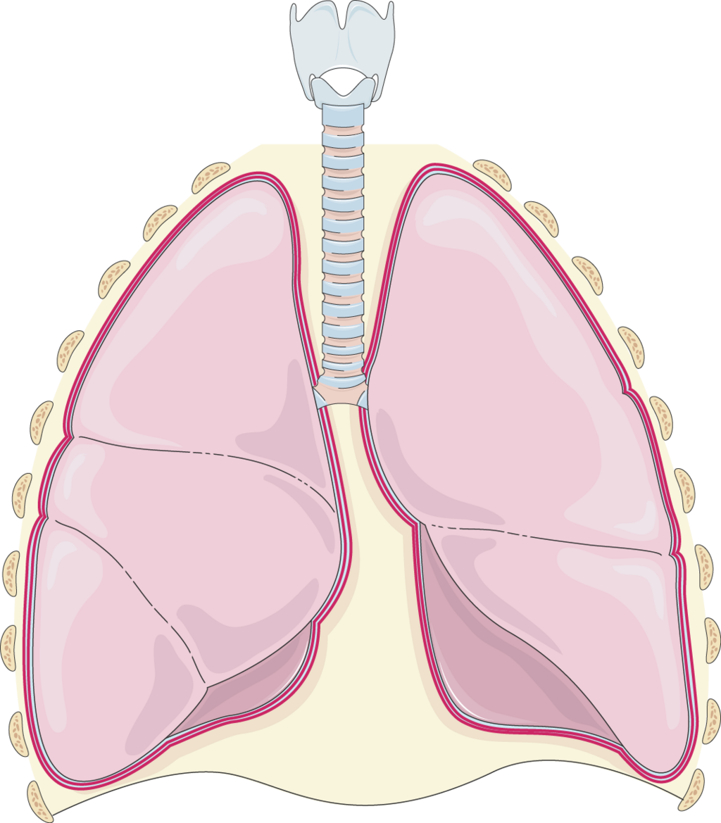
The lungs aren't just floating freely in your chest. They are encased in a two-layered membrane called the pleura. The inner layer, the visceral pleura, covers the surface of the lungs. The outer layer, the parietal pleura, lines the inside of the chest wall.
Between these two layers is a very thin space called the pleural cavity. This cavity contains a small amount of lubricating fluid. This fluid reduces friction, allowing the lungs to glide smoothly against the chest wall as they expand and contract with each breath.
From the entry point at the nose to the millions of alveoli deep within the lungs, the respiratory system is an intricate structure perfectly designed for its vital role. Understanding this anatomy is the first step to appreciating how we breathe.