The Marvels of Human Vision
Eye Anatomy
The Eye's Architecture
The human eye is a remarkably complex organ. Let's explore its structure, starting from the outside and working our way in. The first thing you see when you look at an eye is the tough, white outer layer. This is called the sclera.
Sclera
noun
The white, tough outer layer of the eyeball. It provides structural support and protection for the inner parts of the eye.
Covering the front of the eye is a transparent dome called the cornea. It acts like a window, allowing light to enter. The cornea also does most of the heavy lifting when it comes to focusing that light. A thin, clear membrane called the conjunctiva covers the sclera and lines the inside of your eyelids, helping to keep the eye moist and protected from debris.
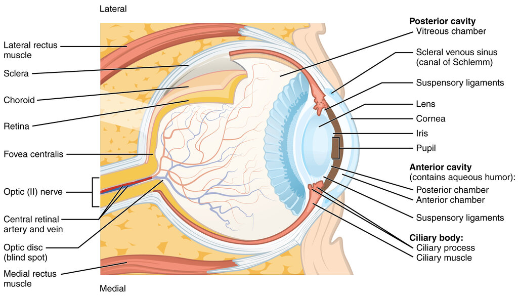
Behind the cornea is the iris, the colored part of the eye. The iris is a muscle that controls the size of the pupil, which is the black dot in the center. The pupil is simply an opening that lets light pass through. In bright light, the iris constricts the pupil to limit the amount of light entering. In dim light, it dilates the pupil to let more light in, just like the aperture of a camera.
Focusing and Filling
Just behind the iris and pupil sits the lens. This transparent, flexible structure fine-tunes the focus started by the cornea. It can change shape to adjust its focusing power, allowing you to see objects clearly whether they are near or far. It flattens for distant objects and becomes more rounded for close ones.
The lens works with the cornea to focus light precisely onto the retina at the back of the eye.
The eye isn't hollow; it's filled with two types of fluid that help it maintain its shape. The space between the cornea and the lens contains a watery fluid called aqueous humor. It provides nutrients to the cornea and lens. The large, central cavity behind the lens is filled with a gel-like substance called vitreous humor. This humor helps support the shape of the eyeball and keeps the retina in place.
The Light-Sensitive Screen
The back of the eye is lined with a thin layer of tissue called the retina. This is where the magic happens. The retina is packed with millions of light-sensitive cells, known as photoreceptors (rods and cones), that convert light into electrical signals. Think of it as the film in a camera or the sensor in a digital camera.
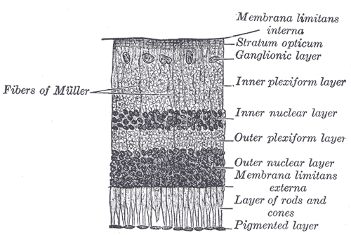
Within the retina is a small, specialized area called the macula. The macula is responsible for our sharp, detailed central vision, which we use for reading, driving, and recognizing faces. At the very center of the macula is a tiny pit called the fovea centralis, or simply the fovea. This area has the highest concentration of cone photoreceptors, which are responsible for color vision and our sharpest daytime sight.
The fovea provides the clearest, most detailed vision. When you look directly at something, you are aiming your fovea at it.
After the retina converts light into electrical signals, these signals travel along the optic nerve to the brain, which interprets them as the images we see.
Now, let's test your knowledge of the eye's anatomy.
What is the primary function of the cornea?
Which part of the eye is a muscle that adjusts the size of the pupil?
Understanding these individual components is the first step in appreciating the complex process of vision.