The Marvel of Human Vision
Introduction to Eye Anatomy
The Eye's Architecture
The human eye is an intricate organ, working much like a sophisticated camera. It gathers light from the world around you, focuses it, and converts it into electrical signals that your brain can interpret as images. To understand how we see, we first need to look at the parts that make this process possible. Let's start with a map of the eye.
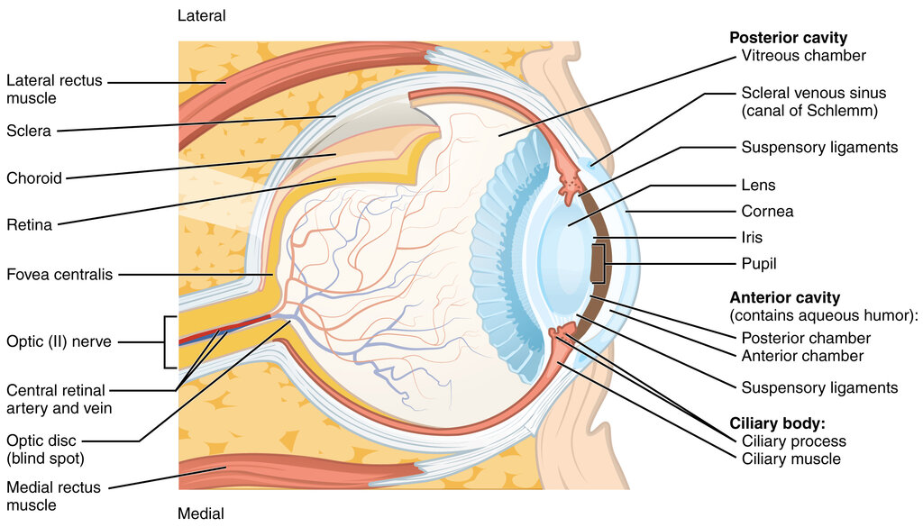
The eye's outermost layer provides both protection and the initial step in focusing light. The tough, white part of the eye is called the sclera. It's a durable, fibrous tissue that covers the entire eyeball except for the very front. Think of it as the eye's protective casing.
At the front, the sclera gives way to a transparent, dome-shaped window called the cornea. The cornea is completely clear, allowing light to enter the eye. It also performs a crucial job: it bends, or refracts, the incoming light, handling most of the eye's focusing power.
Controlling the Light
Once light passes through the cornea, it enters a chamber filled with a watery fluid called the aqueous humor. This fluid helps maintain pressure inside the eye and provides nutrients to the cornea and lens.
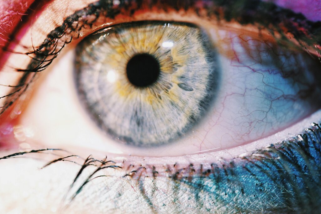
Behind the aqueous humor is the iris, the colored part of your eye. The iris is a muscle that can expand and contract, changing the size of the opening at its center. That opening is the pupil. By adjusting the pupil's size, the iris controls exactly how much light gets deeper into the eye. In bright light, the iris constricts the pupil to a small dot. In dim light, it opens it wide to let as much light in as possible, just like the aperture on a camera.
Iris
noun
The pigmented, muscular part of the eye that surrounds and controls the size of the pupil.
Focusing the Image
Directly behind the pupil sits the lens, a clear, flexible structure. The lens's job is to fine-tune the focus that the cornea started. It can change its shape to adjust for objects at different distances.
This change in shape is controlled by the ciliary muscles, which are attached to the lens. When you look at something far away, these muscles relax, causing the lens to flatten. When you focus on a nearby object, the ciliary muscles contract, allowing the lens to become thicker and more rounded. This adjustment ensures that the light focuses perfectly on the back of the eye.
Past the lens, the main body of the eyeball is filled with a clear, jelly-like substance called the vitreous humor. This gel helps the eye maintain its round shape and is mostly made of water. It provides a clear path for light to travel from the lens to the back of the eye.
The final destination for light inside the eye is the retina. The retina is a thin layer of tissue that lines the back of the eye on the inside. It's light-sensitive and acts like the digital sensor in a camera.
When the focused light hits the retina, specialized cells convert the light energy into electrical signals. These signals are then sent to the brain through the optic nerve for processing. The retina is where the act of seeing truly begins.
Which part of the eye is responsible for the initial and most significant bending of light as it enters?
The iris controls the size of the pupil. This function is most similar to which part of a camera?