The Intricate Anatomy of the Human Eye
External Eye Structures
The Eye's First Defense
The external structures of the eye form a sophisticated barrier, protecting the delicate optical components within. This protective system involves the eyelids, the conjunctiva, and the lacrimal apparatus, each playing a critical role in shielding the eye from harm and maintaining a healthy ocular surface.
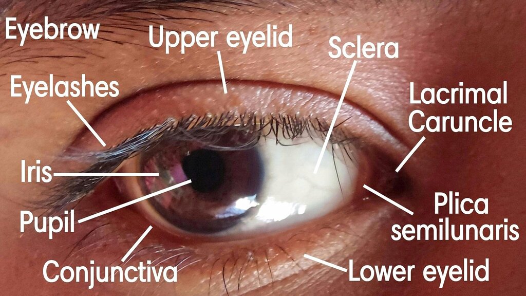
The eyelids, or palpebrae, are more than just skin folds. They are complex structures that provide a physical shield against foreign bodies, excessive light, and injury. Blinking spreads tears across the eye's surface, keeping it moist and clear, while also sweeping away debris.
Structurally, each eyelid is composed of several layers: thin skin on the outside, a layer of muscle, a fibrous tarsal plate, and a specialized mucous membrane called the conjunctiva on the inside. The tarsal plates give the eyelids their shape and firmness. Embedded within these plates are meibomian glands, which secrete an oily substance that forms the outer layer of the tear film. This lipid layer is crucial; it prevents the watery part of the tears from evaporating too quickly.
The Conjunctiva
Lining the inside of the eyelids and covering the white part of the eye (the sclera) is the conjunctiva. This thin, transparent membrane is a single continuous sheet, creating a pocket known as the conjunctival sac. It's impossible for a contact lens to get lost 'behind' your eye because of this barrier.
The conjunctiva is divided into two parts: the palpebral conjunctiva, which lines the eyelids, and the bulbar conjunctiva, which covers the sclera. Rich in blood vessels and containing specialized goblet cells, its primary functions are lubrication and defense. The goblet cells secrete mucin, a key component of the tear film that helps tears adhere to the eye's surface. It also houses lymphoid tissue, which acts as an immunological barrier against pathogens.
The Lacrimal System
Constant moisture is essential for the eye. This is managed by the lacrimal apparatus, a network of glands and ducts responsible for producing and draining tears. The process starts with the lacrimal gland, located in the upper, outer region of the orbit.
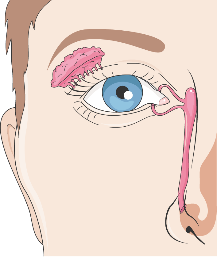
This gland continuously secretes a tear solution—a complex mixture of water, electrolytes, proteins, and antibacterial enzymes like lysozyme. Each time we blink, the eyelids spread this tear film evenly across the cornea.
After lubricating the eye, the tears need to drain. They collect at the inner corner of the eye and enter two tiny openings called the lacrimal puncta, one on the upper and one on the lower eyelid margin. From the puncta, tears flow through small tubes called canaliculi into the lacrimal sac. The sac then drains into the nasolacrimal duct, which carries the tears into the nasal cavity. This connection is why crying often leads to a runny nose.
Together, these external structures form an integrated system. The eyelids provide mechanical protection, the conjunctiva offers a lubricated and immunologically active barrier, and the lacrimal apparatus ensures the ocular surface is constantly cleansed and moisturized. This elegant design keeps our eyes safe and functioning optimally in a world full of potential hazards.
What is the primary function of the meibomian glands located in the eyelids?
The conjunctiva is a continuous membrane that creates a barrier, making it impossible for a contact lens to become lost behind the eye.