Stress Physiology and Vision The Biology of Central Serous Retinopathy
Choroidal Vascular Pathophysiology
The Choroid Under Pressure
You already know how the hypothalamic-pituitary-adrenal (HPA) axis responds to stress by releasing cortisol and catecholamines like epinephrine. Now let's connect that systemic response directly to the eye. The choroid, the vascular layer nestled between the retina and the sclera, is uniquely sensitive to these stress hormones. It's not a passive bystander; it's a primary target.
Chronic elevation of cortisol and catecholamines disrupts the choroid's delicate ability to regulate its own blood flow, a process called autoregulation. Instead of maintaining a stable environment for the retina, the choroidal vessels begin to malfunction. This leads to a condition known as choroidal hyperpermeability, where the vessel walls become excessively leaky. This is the first critical step in the development of Central Serous Retinopathy (CSR).
Chronic sympathetic activation and glucocorticoid release have a large number of deleterious effects.
Think of it as a plumbing system suddenly subjected to fluctuating pressure and corrosive agents. The pipes can't handle the strain. For the choroid, this strain manifests as physical changes to its structure.
Pachychoroid: Thick and Leaky
Sustained high levels of cortisol directly remodel the choroid, leading to a state called 'pachychoroid'. This is characterized by two main features: a significant thickening of the choroid itself and the dilation of the large veins within it, which are then called 'pachyvessels'.
This isn't just a benign swelling. The dilation of these large vessels puts pressure on the overlying choriocapillaris, the fine capillary network responsible for nourishing the outer retina. This pressure, combined with the hormonal effects, causes the choriocapillaris to become congested and hyperpermeable. Fluid begins to seep out of these capillaries, pooling in the space just beneath the retinal pigment epithelium (RPE).
The Molecular Breakdown
This isn't just a mechanical process. At the molecular level, cortisol orchestrates a cascade of events. First, it induces the expression of adrenergic receptor genes within the choroidal vasculature. This makes the vessels hypersensitive to circulating catecholamines like epinephrine, causing them to constrict and dilate erratically.
More importantly, cortisol triggers an increase in a protein called Angiopoietin-like 4 (ANGPTL4). This protein's primary job in this context is to disrupt the integrity of endothelial junctions, the 'glue' that holds the cells of the capillary walls together. As ANGPTL4 levels rise, these junctions weaken and create gaps.
Combined with the increased pressure from the pachyvessels, these new gaps in the choriocapillaris wall provide a direct path for fluid to leak out and accumulate in the sub-retinal space. This is the physiological 'first hit' that initiates the retinal detachment characteristic of CSR.
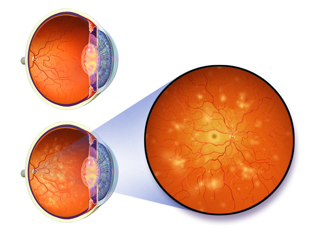
Compounding Factors
The compromised state of the choroid also leads to areas of blood flow stasis and mild ischemia, where parts of the tissue don't receive enough oxygen. This further damages the vessel walls and exacerbates leakage.
Systemic conditions like hypertension add another layer of complexity. A healthy choroid can autoregulate to handle normal fluctuations in blood pressure. However, in a system already weakened by chronic cortisol and catecholamine exposure, the force of high blood pressure can easily overwhelm this autoregulation. This pushes more fluid through the leaky choriocapillaris, worsening the fluid accumulation under the retina and accelerating the progression of CSR.
What is the primary initial effect of chronically elevated cortisol and catecholamines on the choroid's blood vessels?
The term 'pachychoroid' is characterized by which two main features?
Understanding these specific vascular changes is key to seeing how a systemic stress response translates into a focused, sight-threatening condition.