Spinal Rehabilitation and Strengthening
Spinal Anatomy
The Spine's Five Regions
Your spine is more than just a stack of bones. It's a complex, dynamic column that supports your body, protects your spinal cord, and allows for an incredible range of motion. It's divided into five distinct regions, each with its own unique structure and function.
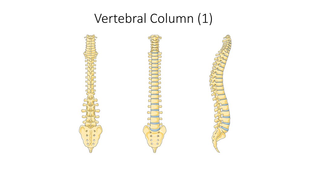
Let's start at the top.
Cervical Spine (C1–C7): This is your neck. It consists of seven small, delicate vertebrae. The cervical spine is the most mobile section, allowing you to turn, tilt, and nod your head. The first two vertebrae are highly specialized. C1, the atlas, holds up the skull, much like the mythological Atlas held up the world. C2, the axis, has a unique peg-like projection called the dens, which the atlas pivots around. This joint is what lets you shake your head "no."
Thoracic Spine (T1–T12): Moving down, we reach the upper and mid-back. The twelve thoracic vertebrae are larger than the cervical ones. Their main job is to anchor the rib cage, forming a protective shield for your heart and lungs. Because of the ribs, this section of the spine is much more rigid and stable, with limited movement.
Lumbar Spine (L1–L5): This is your lower back. The five lumbar vertebrae are the largest and strongest in the spinal column. They have to be, as they bear the majority of your body's weight. This region allows for significant bending and twisting, like when you bend over to pick something up.
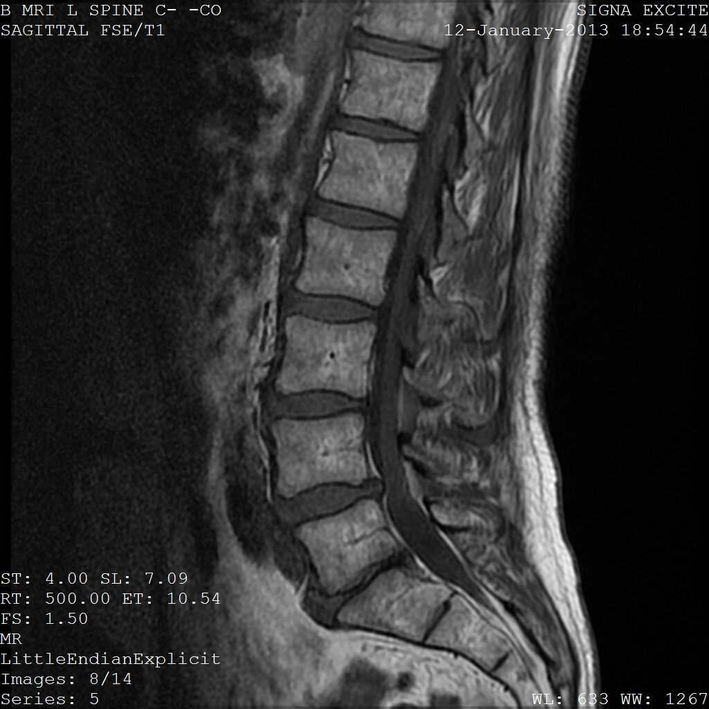
Sacrum and Coccyx: At the base of the spine is the sacrum, a triangular bone formed from five vertebrae that fuse together during adulthood. It connects the spine to the hip bones, transferring weight from the upper body to the legs. Below the sacrum is the coccyx, or tailbone, made of three to five small, fused vertebrae. It serves as an attachment point for various muscles, ligaments, and tendons.
The Spine's Building Blocks
To understand how the spine works as a whole, we need to look at its individual components: the vertebrae that stack up, the discs that cushion them, and the ligaments that hold everything together.
Vertebra
noun
One of the series of bones forming the backbone, having several projections for articulation and muscle attachment, and a hole through which the spinal cord passes.
While vertebrae vary in size and shape depending on the region, they share a basic structure. The main part is the thick, round vertebral body, which is the primary weight-bearing component. Behind it is the vertebral arch, a ring of bone that creates a hollow space. When the vertebrae are stacked, these spaces form the vertebral canal, which houses and protects the delicate spinal cord.
Several bony projections stick out from the arch. The one pointing backward is the spinous process—you can feel these as the bumps along your back. The projections on each side are the transverse processes. These serve as attachment points for muscles and ligaments.
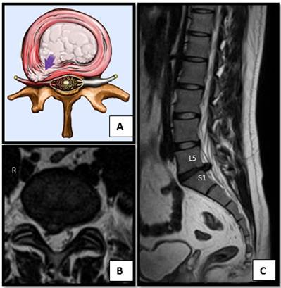
Between each pair of vertebrae lies an intervertebral disc. These are the spine's shock absorbers. Think of them as tiny, durable pillows that cushion the bones during activities like walking, running, and jumping. They also act as pivots that allow the spine to bend and twist.
Each disc has two parts: a tough, fibrous outer ring called the annulus fibrosus and a soft, gel-like center called the nucleus pulposus. The annulus holds the nucleus in place and provides strength, while the jelly-like nucleus provides the cushioning.
Finally, a network of strong, fibrous bands called ligaments holds the entire structure together. They run up and down the spine, connecting vertebrae to each other. The anterior longitudinal ligament runs down the front of the vertebral bodies, while the posterior longitudinal ligament runs down the back. These, along with many smaller ligaments, provide stability, prevent excessive movement, and help protect the spinal cord from injury.
Curves and Posture
When you look at the spine from the side, it's not perfectly straight. It has a series of natural curves that form an S-shape. These curves are essential for balance, flexibility, and shock absorption.
There are two types of curves:
- Lordotic curves: These are the inward curves of the cervical (neck) and lumbar (lower back) regions.
- Kyphotic curves: These are the outward curves of the thoracic (mid-back) and sacral regions.
These curves work together like a coiled spring to absorb shock, maintain balance, and allow the body to move with flexibility. Good posture involves maintaining these natural curves. When you slouch or stand improperly for long periods, you put strain on the muscles and ligaments, which can lead to discomfort and pain over time. Understanding the spine's natural alignment is the first step toward supporting its health.
Now, let's test your knowledge of the spine's structure and function.
Which region of the spine is characterized by its connection to the rib cage, resulting in limited mobility but high stability?
The specialized joint between the atlas (C1) and axis (C2) vertebrae allows for which primary movement?
Understanding the anatomy of your spine is crucial for appreciating how it supports your daily life. From the top of your neck to your tailbone, each component plays a vital role in movement, stability, and protection.