Specific Cardiac Conduction Abnormalities
Sinus Short PR
Decoding the Short PR Interval
In a normal sinus rhythm, the PR interval represents the time it takes for an electrical impulse to travel from the sinus node, through the atria, and down to the ventricles. A standard PR interval is between 120 and 200 milliseconds (ms). When this interval shortens to less than 120 ms, it signifies unusually fast conduction from the atria to the ventricles, a finding that requires careful evaluation.
The PR interval the time taken for the depolarisation to spread from the SA node to the ventricular muscle.
This rapid transit often means the impulse is partially or completely bypassing the atrioventricular (AV) node, which normally acts as a crucial gatekeeper, slowing conduction to allow the ventricles time to fill with blood. But a short PR interval isn't automatically a cause for alarm. Its significance depends entirely on the context and the morphology of the QRS complex that follows.
Benign Anomaly or Red Flag?
A short PR interval can be a normal, benign finding. In some individuals, particularly young athletes, it may simply reflect a highly efficient conduction system. This is often termed (EAVNC), where the AV node itself conducts impulses faster than usual. In these cases, the QRS complex remains narrow (less than 120 ms) and appears normal, as the ventricular activation sequence is unchanged. This pattern was historically referred to as the Lown-Ganong-Levine (LGL) syndrome, though the term is used less frequently today as it describes an ECG pattern rather than a distinct clinical syndrome.
The more concerning cause of a short PR interval is a pre-excitation syndrome, where an accessory pathway—an extra electrical connection outside the normal conduction system—links the atria and ventricles. This bypass tract allows the electrical impulse to circumvent the AV node's delay, leading to premature ventricular activation.
The Telltale Delta Wave
The most well-known pre-excitation syndrome is (WPW). It is characterized by a classic triad on the ECG: a short PR interval (<120 ms), a wide QRS complex (>120 ms), and a distinctive slurring at the beginning of the QRS complex known as a delta wave.
The delta wave represents the early depolarization of the ventricular muscle via the accessory pathway. Because this initial activation doesn't travel through the specialized, high-speed His-Purkinje system, it moves slowly from muscle cell to muscle cell, creating the slurred upslope. The rest of the QRS complex is formed by the fusion of this early activation with the normal activation arriving slightly later through the AV node.
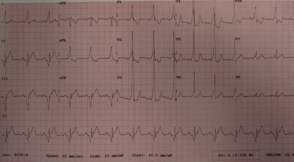
Here's how to distinguish these patterns:
| Feature | Normal Sinus | EAVNC (formerly LGL) | Wolff-Parkinson-White |
|---|---|---|---|
| PR Interval | 120-200 ms | <120 ms | <120 ms |
| Delta Wave | Absent | Absent | Present |
| QRS Width | <120 ms | <120 ms | ≥120 ms |
Clinical Implications
An isolated short PR interval with a normal QRS (EAVNC) is generally considered benign and requires no specific treatment in an asymptomatic person. However, the presence of an accessory pathway, as in WPW, creates a circuit ripe for re-entrant tachycardias. The pathway can conduct impulses both forward (atria to ventricles) and backward (ventricles to atria), setting the stage for arrhythmias.
The most dangerous scenario occurs if a patient with WPW develops atrial fibrillation. In a normal heart, the AV node protects the ventricles by blocking many of the chaotic atrial impulses. In WPW, the accessory pathway can conduct these rapid impulses directly to the ventricles, potentially leading to an extremely fast ventricular rate. This rapid, disorganized ventricular activity can degenerate into ventricular fibrillation, a cause of (SCD).
Because of this risk, even asymptomatic patients with WPW on their ECG are often referred for further evaluation, which may include an electrophysiology study to assess the properties of the accessory pathway and determine the risk of SCD. In contrast, those with a simple short PR interval and no delta wave are typically not at increased risk.
What is the defining time duration for a short PR interval on an ECG?
Which of the following ECG findings is the hallmark of Wolff-Parkinson-White (WPW) syndrome that distinguishes it from a benign short PR interval?
Correctly identifying a short PR interval and distinguishing between its benign and pathological forms is a critical skill in ECG interpretation, ensuring that those at risk receive the appropriate monitoring and care.