Sensorimotor Cortex Motor Imagery EEG
Sensorimotor Cortex Anatomy
The Brain's Action Center
Every move you make, from tapping your finger to walking down the street, starts as a signal in your brain. The region responsible for this is called the sensorimotor cortex. It's not one single spot, but a partnership of two key areas that handle both sending out movement commands and receiving physical sensations from your body.
These two areas sit side by side in the cerebral cortex, the wrinkly outer layer of the brain. They are separated by a prominent groove called the central sulcus. The strip of brain tissue just in front of this groove is the primary motor cortex, while the strip just behind it is the primary somatosensory cortex.
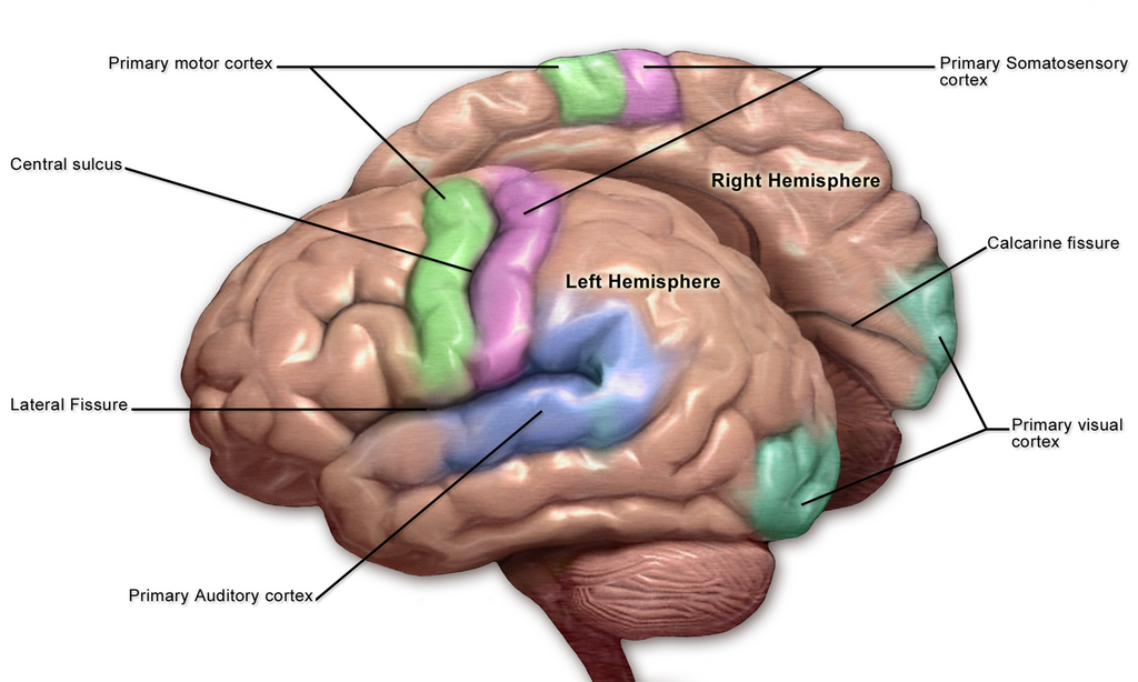
Think of them as a command and feedback system. The motor cortex sends out the orders to move, while the somatosensory cortex receives the incoming reports on touch, pressure, temperature, and pain.
Command Central: The Motor Cortex
The primary motor cortex is located in a ridge known as the precentral gyrus. This area is the main departure point for signals that travel down the spinal cord to your muscles, telling them to contract. It's responsible for executing voluntary movements. When you decide to pick up a cup, the motor cortex generates the precise signals needed to control your arm and hand muscles.
The precentral gyrus houses the primary motor cortex, which initiates voluntary muscle movements.
Sensing the World: The Somatosensory Cortex
Just across the central sulcus lies the primary somatosensory cortex, located in the postcentral gyrus. This region is the main receiving area for sensory information from the skin and internal organs. When you touch a hot surface, receptors in your skin send a signal up to your brain, and it's the somatosensory cortex that processes this information as the sensation of heat and pain.
The postcentral gyrus contains the primary somatosensory cortex, which processes bodily sensations like touch, temperature, and pain.
This constant stream of sensory feedback is crucial for coordinated movement. As you pick up that cup, your somatosensory cortex tells your motor cortex how much pressure your fingers are applying, allowing you to grip it firmly without crushing it.
The Body Map in Your Brain
One of the most fascinating aspects of the sensorimotor cortex is its organization. The neurons in these areas are arranged in a way that maps out the entire body. This is called somatotopic organization. If you could look at the surface of the motor or sensory cortex, you would find a specific spot dedicated to the toes, right next to a spot for the ankle, which is next to the leg, and so on.
But this map isn't a perfect, to-scale representation of the body. Instead, the amount of brain area dedicated to a body part is proportional to how sensitive it is or how much fine motor control it requires. Your lips, tongue, and hands have huge representations in the cortex because they are extremely sensitive and capable of very complex movements. In contrast, your back and thighs have much smaller representations.
This distorted map is often visualized as a character called a homunculus, which means "little person" in Latin. A motor homunculus has enormous hands and a large mouth, reflecting the brain power needed for intricate hand movements and speech. A sensory homunculus is similar, emphasizing the sensitivity of the same areas.
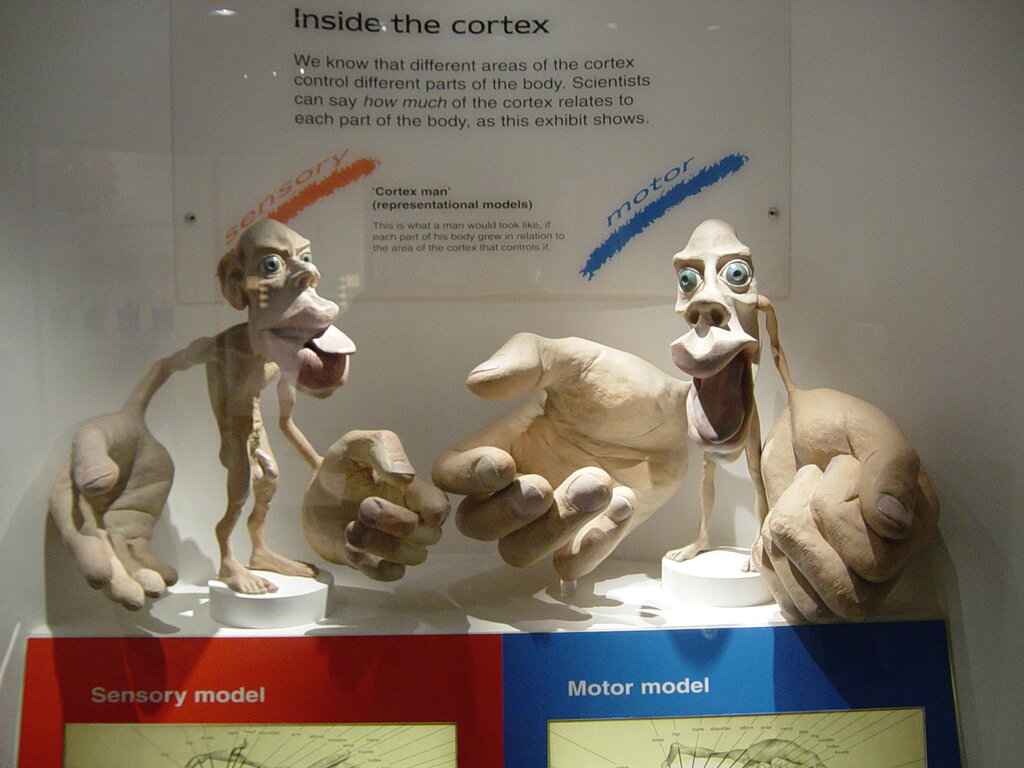
This organization explains why a small paper cut on your finger feels so much more intense than a larger scrape on your back. Your brain simply devotes more processing power to sensations from your fingertips.
Ready to test your knowledge?
What are the two primary areas that form the sensorimotor cortex?
The primary motor cortex is located in the precentral gyrus, while the primary somatosensory cortex is located in the ___________.
Understanding the layout of the sensorimotor cortex provides a clear picture of how our brain dedicates specific resources to controlling our body and perceiving the world through touch.