SEM and TEM Comparison
Electron Matter Interactions
When Electrons Hit Matter
In electron microscopy, an image is formed not by light, but by a focused beam of electrons. The magic happens when this beam strikes a specimen. The electrons interact with the atoms in the sample, producing a variety of signals. By detecting these signals, we can construct a highly magnified image that reveals a world far beyond what our eyes can see.
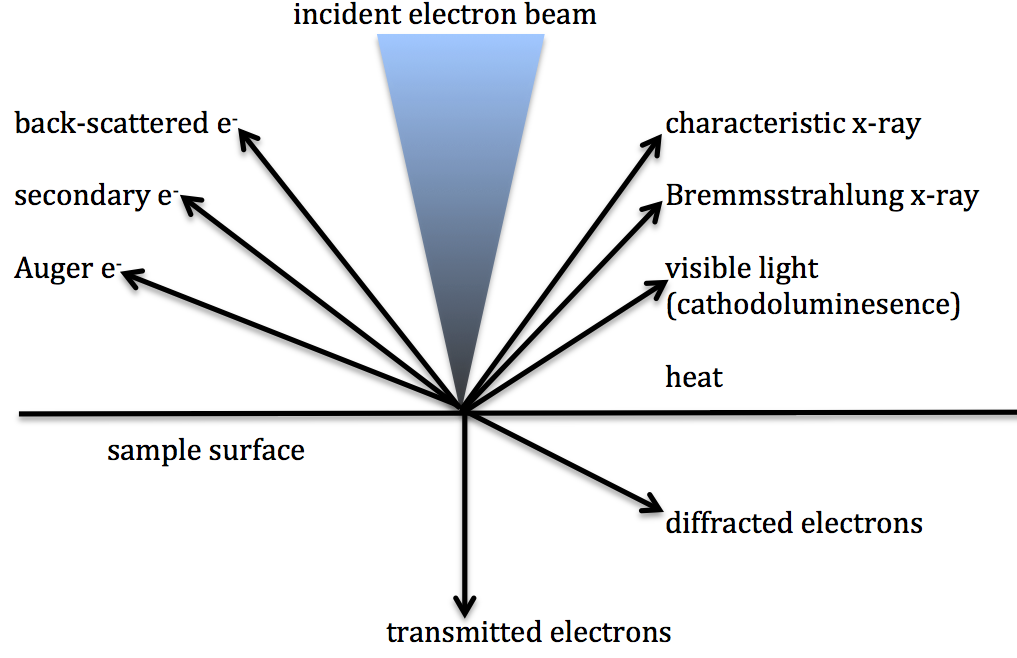
These interactions can be broadly sorted into two types: elastic and inelastic scattering. In an elastic scattering event, an electron from the primary beam changes direction after interacting with an atom, but loses a negligible amount of energy. Think of it like a perfect billiard ball collision. In inelastic scattering, the electron transfers a significant amount of its kinetic energy to the sample's atoms, often by ejecting one of the atom's own electrons. This is more like a bowling ball hitting a pin; energy is transferred, and something new (a secondary electron) is set in motion.
Scanning the Surface
Scanning Electron Microscopy (SEM) is primarily concerned with signals that come from the surface of a specimen. The electron beam scans across the sample, and detectors pick up the electrons that fly off. The two most important signals for SEM imaging are backscattered electrons and secondary electrons.
Backscattered electrons (BSE) are high-energy electrons from the primary beam that have undergone elastic scattering with atomic nuclei in the sample. They essentially "bounce off" the atoms and fly back out towards the detector. The likelihood of this happening is strongly related to the atom's atomic number (Z). Heavier elements, with more protons in their nucleus, are much more effective at scattering electrons back. This makes BSE imaging an excellent tool for compositional analysis; regions with heavier elements appear brighter in the image.
Secondary electrons (SE) are a product of inelastic scattering. When a high-energy primary electron passes near an atom, it can transfer enough energy to knock one of the atom's own loosely bound electrons out of its orbit. These ejected electrons are the secondary electrons and have very low energy. Because of their low energy, only SEs generated very close to the surface (typically within a few nanometres) can escape to be detected. This makes SE imaging extremely sensitive to surface topography. Nooks, crannies, and sharp edges all produce a high yield of secondary electrons, resulting in detailed, three-dimensional-looking images.
Seeing Through the Sample
Transmission Electron Microscopy (TEM) works differently. Instead of looking at electrons that bounce off the surface, TEM analyzes the electrons that pass through a very thin specimen. The sample must be incredibly thin, often less than 100 nanometres, to allow electrons to penetrate it. The resulting image is a 2D projection of the sample's internal structure.
As the electron beam passes through the specimen, a few things can happen:
- Unscattered Electrons: Some electrons pass straight through without interacting with any atoms at all. These are also called direct beam electrons.
- Elastically Scattered Electrons: These electrons are deflected by atoms in the sample but retain their energy. They are crucial for forming diffraction patterns, which give information about the crystalline structure of the material.
- Inelastically Scattered Electrons: These electrons lose energy as they interact with the sample's electrons. They carry information about the elemental composition and chemical bonding within the sample. While useful for certain analytical techniques, they can create a type of blur called chromatic aberration in standard TEM images.
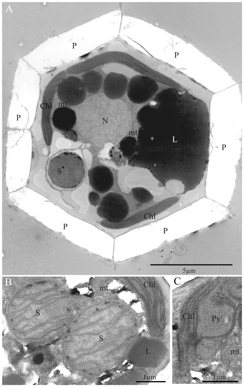
In a standard TEM bright-field image, an aperture is used to block most of the scattered electrons. The image is formed primarily by the unscattered electrons. Therefore, areas of the sample that are thicker or denser (containing heavier atoms) will scatter more electrons away from the aperture and appear darker in the final image. This is the fundamental source of contrast in bright-field TEM imaging.
In Scanning Electron Microscopy (SEM), which signal is most sensitive to surface topography, revealing fine details like edges and textures?
An SEM image created using a backscattered electron (BSE) detector shows two distinct regions: one bright and one dark. What is the most likely interpretation?
Understanding how electrons interact with a sample is the first step to interpreting the complex images these powerful microscopes produce. Whether you're mapping elements on a surface with BSE or peering inside a cell with TEM, it all comes down to scattering.