SEM and TEM Comparison
Electron Optics Refresher
Beyond the Limits of Light
To see smaller things, you need a smaller ruler. In microscopy, the 'ruler' is the wavelength of the illumination source. Light microscopy hits a wall because the wavelength of visible light is relatively long, typically 400 to 700 nanometres. You simply can't use a metre stick to measure a millimetre accurately.
Enter the electron. Thanks to wave-particle duality, electrons, which we often think of as particles, also behave like waves. The crucial difference is that their wavelength is far, far shorter than that of light. This is governed by the de Broglie relationship.
An electron accelerated by 100,000 volts has a wavelength of about 0.004 nanometres. Compare that to the hundreds of nanometres for visible light. This dramatically shorter wavelength is what gives electron microscopes their extraordinary resolving power, allowing us to visualise structures at the atomic scale.
Shorter wavelength equals higher resolution. It's the fundamental advantage of using electrons instead of photons.
Generating the Beam
The journey begins at the electron gun, the source of the electron beam. Its job is to emit a steady, high-intensity stream of electrons and accelerate them down the microscope column. The type of gun used significantly impacts the microscope's performance, particularly the brightness and coherence of the beam. There are two main families of electron sources.
| Emission Type | How it Works | Pros | Cons |
|---|---|---|---|
| Thermionic | A tungsten filament or LaB₆ crystal is heated to over 2500 K, 'boiling' electrons off its surface. | Robust, inexpensive, less stringent vacuum requirements. | Lower brightness, larger spot size, shorter lifespan. |
| Field Emission | A very strong electric field is applied to a sharp tungsten tip, pulling electrons out via quantum tunnelling without extreme heat. | Much higher brightness, smaller spot size, longer lifespan. | Expensive, requires an ultra-high vacuum, less stable. |
A brighter source provides a stronger signal, which is crucial for high-resolution imaging and analysis. Field Emission Guns (FEGs) produce a beam that is up to 1,000 times brighter than a thermionic source, making them the standard for high-performance TEM and SEM systems.
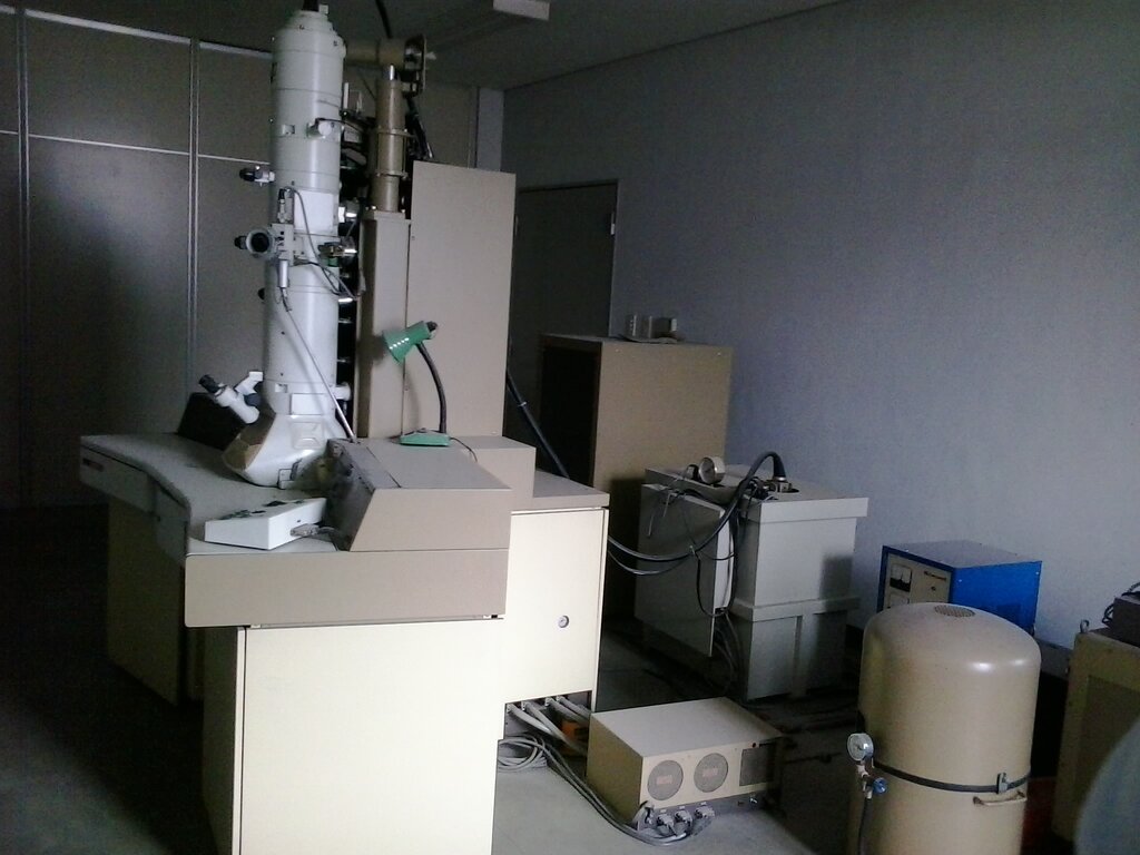
Focusing with Fields
Once the electrons are generated and accelerated, they must be controlled. Since electrons are charged particles, they can be deflected by magnetic fields. This is the principle behind electromagnetic lenses, which perform the same function for an electron beam that glass lenses do for light.
An electromagnetic lens is essentially a coil of wire wrapped around a soft iron core. When current flows through the coil, it generates a precise, rotationally symmetric magnetic field within the lens's gap. As electrons pass through this field, they experience a force that pushes them towards the central axis. The strength of the lens, and thus its focal length, can be changed almost instantly by adjusting the current in the coil.
The illumination system of both SEM and TEM instruments uses a series of these lenses. Condenser lenses shape the beam and control the amount of current that hits the sample, similar to a tap controlling water flow. The objective lens is the most critical component, as it forms the final, focused probe of electrons that interacts with the specimen. The quality of this lens is paramount for achieving high resolution.
However, just like glass lenses, electromagnetic lenses are not perfect. They suffer from defects called that can blur the image. The most significant of these is spherical aberration, where electrons travelling on the outer edges of the lens are focused more strongly than those near the centre. This causes a sharp point to be imaged as a blurry disc. Astigmatism is another common issue, where the lens has different focal lengths for different directions, stretching a point into a line. Modern microscopes use complex lens designs and correctors to minimise these effects.
Why do electron microscopes have a higher resolving power than light microscopes?
What is the most significant lens aberration that causes electrons on the outer edges of a lens to be focused more strongly than those near the centre?