Selling Advanced Cancer Diagnostics to Oncologists
Understanding Tissue-Based Diagnostics
Getting the Sample
When doctors suspect cancer, they often need to examine a piece of the suspicious tissue directly. This process of removing a small sample for testing is called a biopsy. It's the cornerstone of cancer diagnosis.
A biopsy provides a physical piece of the puzzle, allowing doctors to look at the cells themselves.
There are several ways to perform a biopsy, depending on the tumor's location. A fine-needle aspiration uses a very thin needle to draw out cells, which is less invasive. A core needle biopsy uses a slightly larger, hollow needle to remove a small cylinder of tissue. For tumors that are hard to reach or need a larger sample, a surgeon might perform a surgical biopsy, removing the entire lump (excisional biopsy) or just a piece of it (incisional biopsy).
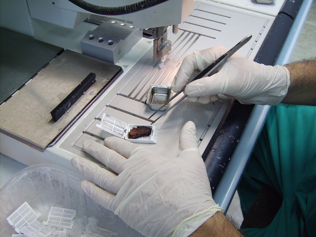
Regardless of the method, the goal is the same: to get a high-quality tissue sample that accurately represents the suspected tumor.
Under the Microscope
Once the sample arrives in the lab, the real investigation begins. This is the realm of histopathology, the study of diseased tissue. A specialist called a pathologist prepares the tissue to be viewed under a microscope.
The sample is preserved in formalin, embedded in a block of paraffin wax, and then sliced into incredibly thin sections—thinner than a human hair. These transparent slices are placed on glass slides and stained with dyes.
The most common stain, Hematoxylin and Eosin (H&E), turns cell nuclei blueish-purple and the rest of the cell pink. This contrast makes the tissue's structure visible.
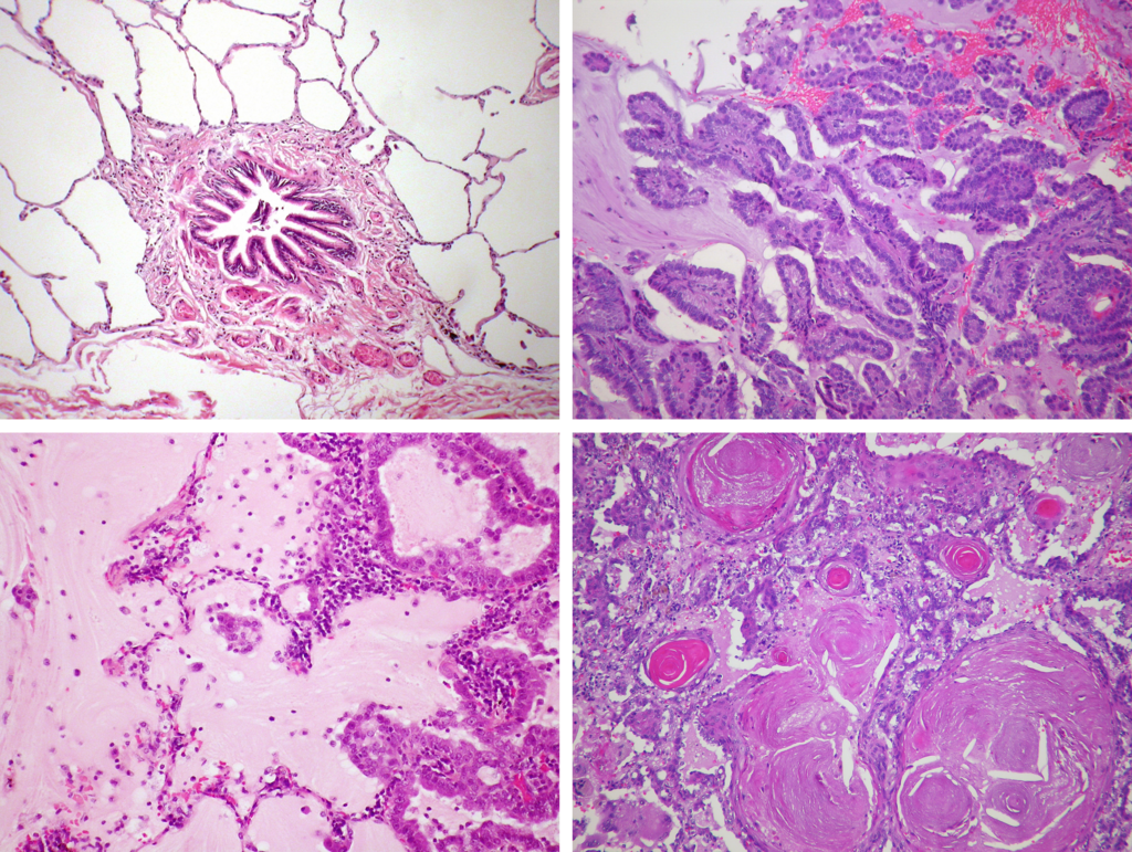
The pathologist examines the stained tissue, looking for the tell-tale signs of cancer. Are the cells disorganized? Are their nuclei unusually large or irregularly shaped? Are they invading surrounding tissues? The answers to these questions help determine if cancer is present, what type it is, and how aggressive it might be. This analysis is crucial for creating a treatment plan.
Staining for Specifics
Sometimes, the appearance of cells isn't enough to make a definitive diagnosis. That's where a technique called immunohistochemistry (IHC) comes in. Think of it as using molecular highlighters.
IHC uses antibodies that are designed to stick to specific proteins on or inside cells. These antibodies are linked to a dye. When applied to the tissue sample, the antibodies bind to their target proteins, causing those specific cells to change color. This allows pathologists to identify cells based on the proteins they produce, which can distinguish between different types of cancer or identify targets for certain drugs.
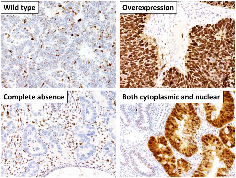
For example, some breast cancers have high levels of a protein called HER2. An IHC test can reveal which cells are overproducing this protein, guiding doctors to use therapies that specifically target HER2.
The Limits of the Biopsy
While tissue biopsies are the gold standard for diagnosis, they aren't perfect. The procedures themselves are invasive, carrying risks like pain, bleeding, and infection. Depending on the tumor's location, such as deep within the lung or brain, a biopsy can be difficult and dangerous to perform.
Another major challenge is tumor heterogeneity. Tumors are not uniform; different parts can have different genetic makeups. A biopsy only samples one small spot, which might not represent the entire tumor.
Imagine a tumor is like a bag of mixed jelly beans. A needle biopsy might pull out only red ones, missing the blue and green ones entirely. This means the initial diagnosis might not capture the full complexity of the cancer, which can change as it grows and spreads.
Finally, because they are invasive, biopsies are not ideal for monitoring a patient's cancer over time. Repeated procedures are often impractical. These limitations have pushed researchers to develop new diagnostic methods that can complement the traditional tissue biopsy.