Science of Recovery and Biological Healing
Inflammation and Cellular Signalling
Coordinating the Response
When tissue is damaged, the body doesn't just patch the hole. It launches a coordinated, multi-stage operation called inflammation. The first step is to manage the immediate crisis. Almost instantly, blood vessels near the injury site change their behaviour. This is the vascular phase.
Imagine a busy motorway. An accident happens, and traffic control needs to reroute vehicles, get emergency services in, and clear the debris. The body does something similar. Injured cells and nearby immune cells release chemical alarms. These signals cause two things to happen: vasodilation, where the blood vessels widen, and increased vascular permeability, where the vessel walls become leaky.
Widening the vessels slows down blood flow, allowing immune cells to pause and take notice of the damage. The leakiness creates exits for these cells and for blood plasma to get to the site of injury.
Chemical Mediators
The entire inflammatory response is directed by a cocktail of chemical signals. Mast cells, which are immune cells stationed in tissues, are among the first to react. They degranulate, releasing a flood of histamine. Histamine is a primary driver of the immediate vasodilation and permeability you see as swelling and redness.
Soon after, other mediators join in. Bradykinin, a peptide formed in the blood, contributes to vasodilation and also has another important job: it makes the area sensitive to pain. This pain response is a protective mechanism, discouraging you from using or touching the injured area. At the same time, enzymes in the damaged tissue begin producing prostaglandins. These lipid compounds amplify the effects of histamine and bradykinin, intensifying the inflammatory response and contributing to fever.
These chemical signals don't just act locally. They are the distress call that summons reinforcements from the bloodstream, initiating the next phase of the operation.
The Cellular Cleanup Crew
With the chemical signals broadcasting, it's time for the cellular phase. The bloodstream is full of white blood cells, or leukocytes, which are the body's mobile defence and repair units. The changes in blood flow and vessel permeability allow these cells to leave the circulation and enter the tissue, a process called leukocyte extravasation.
The first to arrive are the neutrophils. Think of them as the first responders. They are highly mobile and are experts at phagocytosis, which means 'cell eating'. They engulf and digest pathogens, dead cells, and other debris. A neutrophil's life is short and intense; they do their job for a few hours to a few days and then die, becoming a major component of pus.
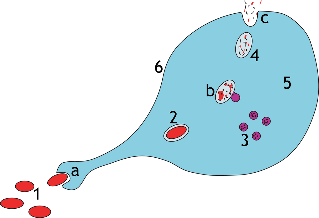
Following the neutrophils are the macrophages. These are the heavy-duty cleanup crew. Macrophages are much larger and live longer than neutrophils. They are voracious phagocytes, clearing away the remaining debris, dead neutrophils, and any lingering pathogens. But their job is more complex than just cleaning up. They are also master regulators of the healing process.
Switching from Defence to Repair
For healing to begin, the inflammation must be resolved. A persistent inflammatory state can cause more harm than good, leading to chronic disease. The key to this transition lies with the macrophages. Initially, macrophages exist in a pro-inflammatory state, known as the M1 phenotype. M1 macrophages are aggressive, producing signals that recruit more immune cells and kill pathogens.
Once the area is clear of threats, local signals instruct the macrophages to change their identity. They switch to an anti-inflammatory M2 phenotype. These M2 macrophages release a completely different set of chemical signals. Instead of calling for battle, these signals promote tissue repair. They encourage the growth of new blood vessels (angiogenesis), stimulate cells called fibroblasts to lay down new extracellular matrix, and generally manage the transition into the proliferative phase of healing.
As healing progresses, anti-inflammatory signals are activated to reduce inflammation and promote tissue repair.
This is a critical control point. If this switch fails, inflammation can become chronic, where the constant presence of M1 macrophages and other immune cells leads to progressive tissue destruction instead of repair.
Let's review the key terms from this section.
Now, test your understanding of the process.
What are the two primary effects caused by chemical alarms like histamine during the initial vascular phase of inflammation?
Which chemical mediator is primarily responsible for making an injured area sensitive to pain, as a protective mechanism?
The resolution of inflammation is an active, highly regulated process. It involves the production of specialised pro-resolving mediators, the departure of immune cells, and the restoration of normal tissue function, paving the way for complete healing.