Restoring Vision Medical Interventions
Eye Anatomy
The Eye's Outer Window
Light's journey into your eye begins at the cornea. This is the transparent, dome-shaped layer that covers the front of your eye, including the iris and pupil. Think of it as the crystal on a watch face—it's a clear, protective shield.
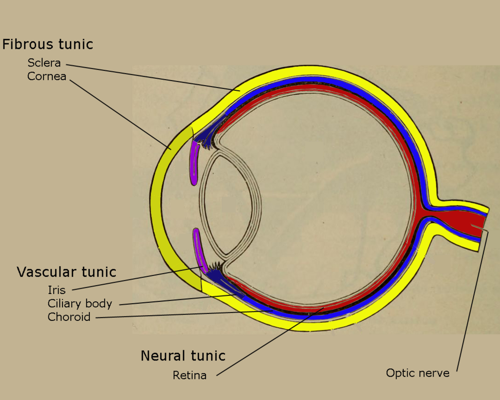
But the cornea does more than just protect. Its curved surface is responsible for most of the eye's focusing power. As light passes through it, the cornea bends, or refracts, the light rays, starting the process of creating a sharp image.
The cornea's primary job is to bend incoming light, performing the initial, heavy-duty focusing for your vision.
The Adjustable Focus
After passing through the cornea and the pupil, light hits the lens. This structure is located just behind the iris (the colored part of your eye). Unlike the cornea, which has a fixed shape, the lens is flexible.
lens
noun
A transparent, biconvex structure in the eye that, along with the cornea, helps to refract light to be focused on the retina.
The lens acts like the autofocus on a camera. Tiny muscles attached to it can contract or relax, changing the lens's curvature. This change allows your eye to fine-tune its focus, letting you see objects clearly whether they are up close or far away. It’s this adaptability that allows you to switch your gaze from a book in your hands to a bird outside your window.
The Screen Inside
Once light is focused, it needs a place to land. That place is the retina, a thin layer of tissue lining the back of the eye. If the eye is a camera, the retina is the digital sensor. It's packed with millions of light-sensitive cells called photoreceptors.
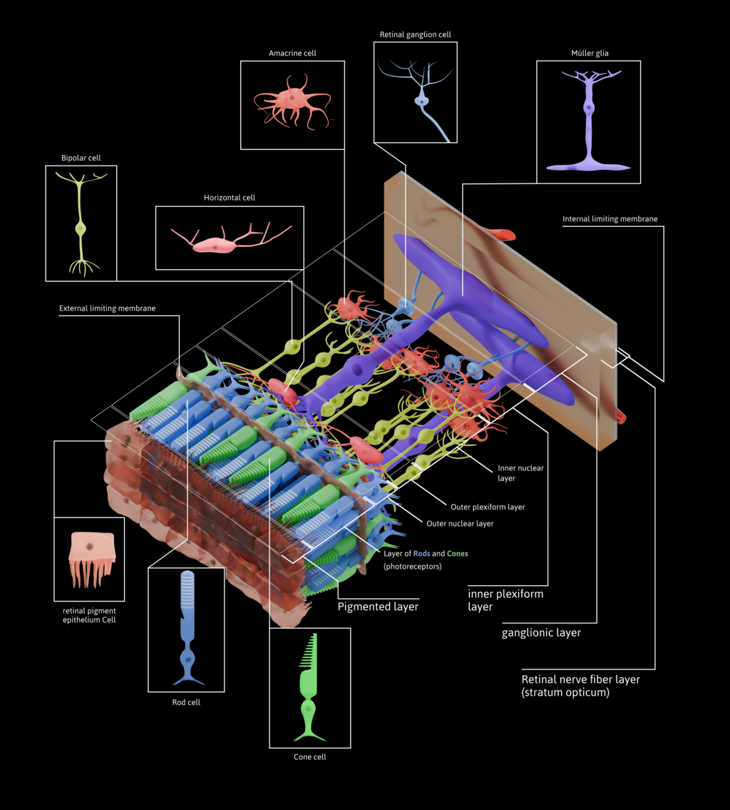
These photoreceptors (rods and cones) do something amazing: they convert the light energy into electrical signals. This is where the physical act of light detection becomes a biological message that your nervous system can understand. The retina doesn't just see light; it translates it into the language of the brain.
The Data Cable and Filling
So, the retina has turned the image into electrical signals. Now what? These signals need to get to the brain for processing. The optic nerve is the high-speed data cable that makes this happen. It's a bundle of more than a million nerve fibers that travels from the back of each eye to the brain.
The optic nerve carries all the visual information from the retina to the brain, which then interprets these signals into the images we perceive as sight.
Finally, the main body of the eyeball isn't empty. It's filled with a clear, gel-like substance called the vitreous humor. This gel helps the eye maintain its round shape, acting like a gentle internal pressure system. It also allows light to pass through unobstructed from the lens to the retina.
Which part of the eye is responsible for the majority of its light-focusing power?
The lens is similar to a camera's autofocus because it can change its shape to see objects clearly at various distances.
Understanding these core parts—cornea, lens, retina, optic nerve, and vitreous humor—gives you a foundational map of how we see.