Renal System for Medical Licensing Exams
Renal Anatomy
The Urinary System's Blueprint
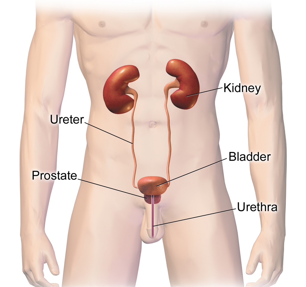
Your urinary system is a highly efficient waste removal and fluid-balancing plant. It's composed of four main parts: two kidneys, two ureters, one bladder, and one urethra. The kidneys are the primary filters, cleaning your blood and producing urine. The ureters are tubes that carry this urine from the kidneys to the bladder, which acts as a temporary storage tank. Finally, the urethra is the tube through which urine exits the body.
A Closer Look at the Kidneys
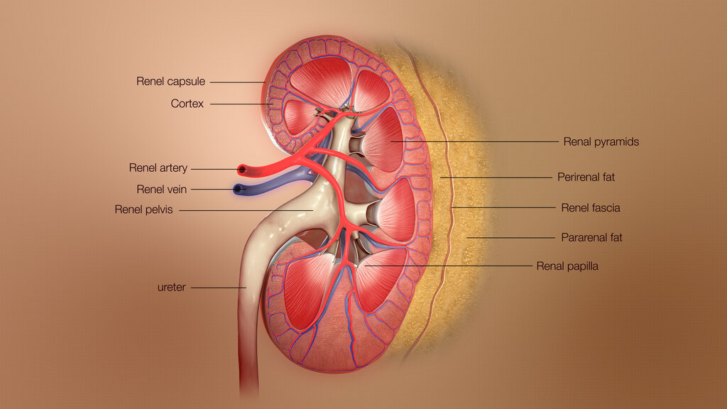
You have two bean-shaped kidneys, each about the size of a fist. They're located on either side of your spine, just below your rib cage, and are protected by a layer of fat. A tough, fibrous layer called the renal capsule covers the outside of each kidney, providing a barrier against injury.
Slicing a kidney open reveals three distinct regions. The outermost layer is the renal cortex, which has a granular appearance because it contains the filtering units of the kidney. Beneath the cortex is the renal medulla, made up of cone-shaped structures called renal pyramids. The innermost region is the renal pelvis, a funnel-shaped area that collects urine from the pyramids and channels it into the ureter.
The Nephron: The Kidney's Workhorse
Each kidney contains about a million microscopic filtering units called nephrons. These are the true powerhouses of the urinary system, responsible for removing waste from the blood. Each nephron has two main parts: a renal corpuscle and a renal tubule.
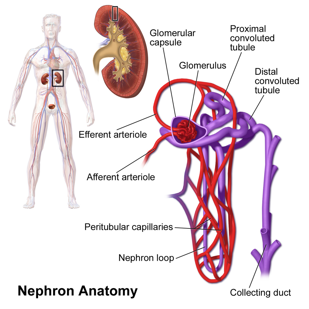
The process begins in the renal corpuscle. This structure consists of the glomerulus, a tangled bundle of tiny capillaries, and the Bowman's capsule, a cup-shaped sac that surrounds it. Blood enters the glomerulus under pressure, forcing water, salts, and waste products out of the blood and into the Bowman's capsule. Larger components like red blood cells and proteins are too big to pass through and remain in the blood.
From the Bowman's capsule, the filtered fluid, now called filtrate, enters the renal tubule. This long, twisting tube is where the fine-tuning happens. It has three main sections:
Proximal Convoluted Tubule (PCT): The first section, located in the cortex, where most of the reabsorption of useful substances like glucose, vitamins, and water occurs.
Loop of Henle: A long, hairpin-shaped loop that dips down into the medulla. Its primary role is to create a concentration gradient, which helps conserve water.
Distal Convoluted Tubule (DCT): The final section of the tubule, also in the cortex, where more selective reabsorption and secretion take place to precisely regulate the body's electrolyte and pH balance.
After passing through the renal tubule, the fluid is officially considered urine. It then flows into a collecting duct, which gathers urine from several nephrons and carries it through the renal pyramids to the renal pelvis.
The Pathway Out
Once urine leaves the kidneys, it travels down the ureters. These are two narrow, muscular tubes that connect each kidney to the bladder. The walls of the ureters contain smooth muscle that contracts in waves to push urine downward, away from the kidneys.
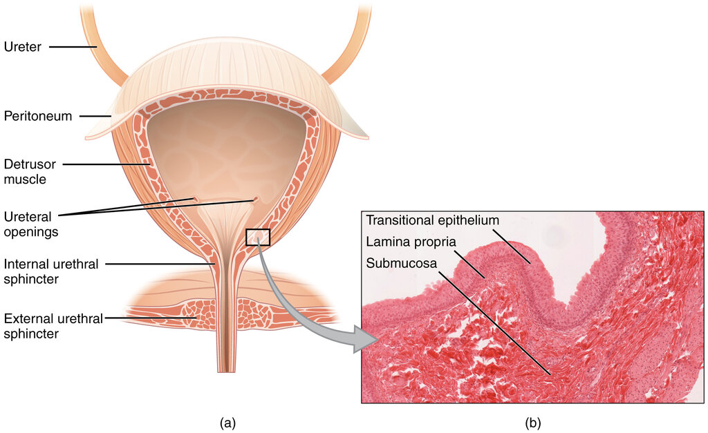
The bladder is a hollow, muscular, and stretchable organ that sits in your pelvic cavity. Its main job is to store urine. The bladder's inner lining is made of transitional epithelium, a special type of tissue that allows it to expand significantly without tearing as it fills.
When the bladder is full, nerve signals are sent to the brain, creating the urge to urinate. During urination, the bladder muscle contracts, and a sphincter muscle at the bladder's exit relaxes, allowing urine to flow into the urethra. The urethra is the final tube that carries urine from the bladder out of the body. Its structure differs between males and females, being much shorter in females.
Ready to check your understanding of the urinary system's anatomy?
What is the correct sequence for the flow of urine out of the body?
The microscopic filtering units located in the kidneys are called _______.
Now you have a complete map of the urinary system's structures, from the large-scale organs down to their microscopic functional units.