Radiography for Cholecystitis
Gallbladder Anatomy
The Gallbladder's Place in the Body
The gallbladder is a small, pear-shaped organ situated in a shallow depression on the undersurface of the liver. It's typically about 7 to 10 centimeters long and is located in the upper right quadrant of your abdomen. Think of it as a small pouch tucked away for safekeeping, right beneath the body's largest internal organ.
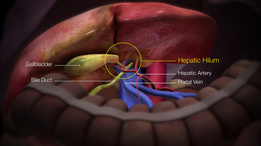
Its primary job is to store and concentrate bile, a digestive fluid produced by the liver. When you eat fatty foods, the gallbladder contracts, squeezing this bile out into the small intestine to help with digestion.
Anatomically, the gallbladder is divided into three parts:
- Fundus: The rounded, bottom end that often peeks out from under the liver's edge.
- Body: The main, central portion.
- Neck: The narrow, tapering end that connects to the cystic duct.
Wall Structure and Layers
The gallbladder wall isn't just a simple sac; it's composed of several distinct layers that enable it to do its job. From the inside out, these layers are the mucosa, muscularis, and serosa.
The innermost layer, the mucosa, has many folds called rugae. These folds flatten out as the gallbladder fills with bile, allowing it to expand like a small balloon.
Just outside the mucosa is the muscularis, a layer of smooth muscle. When stimulated by hormones, this muscle contracts forcefully to eject bile into the duct system. The outermost layer is the serosa, a thin membrane that covers the surface of the gallbladder that is not in direct contact with the liver. The part attached to the liver is covered by a connective tissue layer called the adventitia.
The Biliary Highway
The gallbladder doesn't work in isolation. It’s a key part of the biliary system, a network of ducts that transports bile. The journey begins in the liver, where bile is produced. It flows through tiny right and left hepatic ducts, which merge to form the common hepatic duct.
Bile can then flow down the common hepatic duct to be stored in the gallbladder via the cystic duct. When needed, the bile is released back through the cystic duct, which joins the common hepatic duct to form the common bile duct. This final duct carries bile down to the first part of the small intestine, the duodenum, where it mixes with food.
Blood Supply and Nerves
Like any organ, the gallbladder needs a dedicated blood supply to function. The cystic artery, which usually branches off the right hepatic artery, is the main vessel that supplies the gallbladder with oxygen-rich blood. Deoxygenated blood is drained away through cystic veins.
The gallbladder's functions are also regulated by nerves. The nervous system controls the contraction of the muscle layer and can also transmit pain signals. Nerves from both the celiac plexus and the vagus nerve connect to the gallbladder, coordinating its activity with the rest of the digestive system.
What is the primary function of the gallbladder?
Which of the following lists the three anatomical sections of the gallbladder?