Physiology of the Urinary System
Renal Filtration Dynamics
The Filtration Barrier
Your blood is cleaned inside the nephron's renal corpuscle, a process that begins with a highly selective filter. This isn't a simple sieve; it's a sophisticated, three-layered biological membrane designed to let water and small solutes pass from the blood into the Bowman's capsule while holding back blood cells and large proteins like albumin.
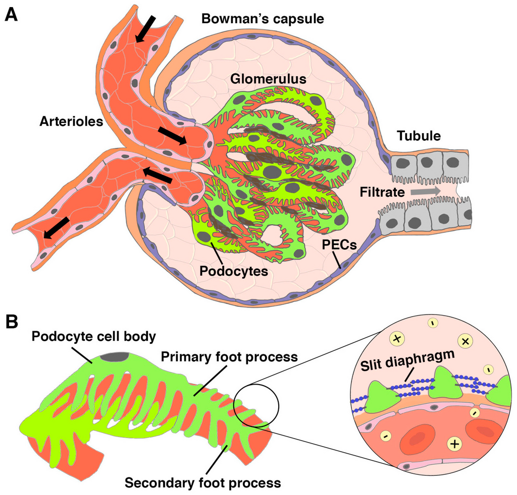
The first layer is the fenestrated endothelium of the glomerular capillaries. These cells are dotted with pores that block blood cells but allow all other plasma components through.
Next is the glomerular basement membrane (GBM), a gel-like layer that prevents the filtration of larger proteins. It's also negatively charged, which repels negatively charged proteins, adding a layer of electrostatic selectivity to the physical barrier.
Finally, specialized cells called podocytes wrap around the capillaries. They have long, finger-like extensions called foot processes that interlock, creating narrow gaps known as filtration slits. These slits are the final barrier, preventing the passage of medium-sized proteins that might have slipped through the first two layers.
The Forces of Filtration
The movement of fluid across this barrier is governed by a balance of pressures, collectively known as Starling forces. Filtration only occurs if the net pressure pushes fluid out of the capillaries and into the Bowman's capsule.
The main driving force is the glomerular hydrostatic pressure (GHP), which is the blood pressure within the glomerular capillaries. It's typically higher than in other capillaries in the body, around 55 mmHg, because the efferent arteriole (which exits the glomerulus) is narrower than the afferent arteriole (which enters). This difference in diameter creates a bottleneck, increasing the pressure that pushes fluid out.
Two forces oppose this filtration:
- Capsular hydrostatic pressure (CHP): This is the pressure exerted by the fluid already inside the Bowman's capsule, pushing back against the capillary. It's usually about 15 mmHg.
- Blood colloid osmotic pressure (BCOP): This is the "pull" exerted by proteins, primarily albumin, that remain in the blood. Since these proteins can't pass through the filter, they create an osmotic gradient that draws water back into the capillaries. This pressure is typically around 30 mmHg.
These forces combine to determine the Net Filtration Pressure (NFP), the overall pressure driving filtration.
Using our typical values, the NFP would be . This positive pressure, though small, is enough to drive the filtration of about 180 liters of fluid from the blood each day.
Keeping Filtration Stable
The body needs a relatively constant Glomerular Filtration Rate (GFR) to maintain homeostasis, even when your systemic blood pressure fluctuates—for example, during exercise or rest. The kidneys have remarkable intrinsic mechanisms to autoregulate GFR over a wide range of blood pressures (typically 80-180 mmHg mean arterial pressure).
One key mechanism is the myogenic response of the afferent arteriole. The smooth muscle in the arteriole's wall contracts when it's stretched by high blood pressure. This constriction reduces blood flow into the glomerulus, lowering GHP and keeping GFR stable. Conversely, if blood pressure drops, the arteriole dilates, increasing blood flow and maintaining GFR.
A more complex mechanism is tubuloglomerular feedback, which involves a specialized structure called the juxtaglomerular apparatus (JGA).
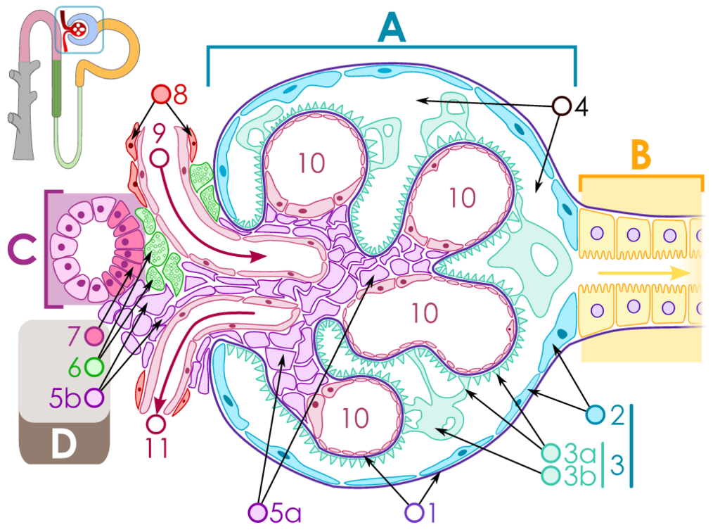
The JGA includes a group of cells in the distal convoluted tubule called the macula densa. These cells act as sensors, monitoring the flow rate and the concentration of sodium chloride (NaCl) in the filtrate.
If GFR increases, fluid flows faster through the tubule, leaving less time for NaCl to be reabsorbed. The macula densa cells detect this higher NaCl concentration and release signaling molecules (like ATP). These signals cause the adjacent afferent arteriole to constrict, reducing blood flow into the glomerulus and bringing GFR back down.
If GFR decreases, the opposite happens. Less NaCl is detected, the afferent arteriole dilates, and GFR increases. This elegant feedback loop allows the nephron to fine-tune its own filtration rate.
Which layer of the glomerular filtration membrane is responsible for preventing the passage of large proteins primarily through electrostatic repulsion?
What is the primary force that drives the movement of fluid out of the glomerular capillaries and into the Bowman's capsule?
These filtration dynamics are the first and most critical step in urine formation, ensuring that waste is efficiently cleared while the body's essential components are retained.