PET Scans in Advanced Prostate Cancer Care
PET scan fundamentals
How PET Scans See Inside You
Positron Emission Tomography, or PET, is a type of medical imaging that shows how your organs and tissues are functioning at a cellular level. Unlike an X-ray or CT scan, which reveals anatomical structure, a PET scan highlights metabolic activity. It essentially creates a map of how much energy different parts of your body are using.
This is especially useful in oncology. Cancer cells are notoriously greedy, consuming energy much faster than healthy cells. A PET scan can spot these high-energy areas, helping doctors detect tumors, see if cancer has spread, and check if a treatment is working.
Using Radiotracers
The key to a PET scan is a special substance called a radiotracer. Before the scan, you receive a small, safe injection of this tracer, which then travels through your bloodstream. The tracer is made of two parts: a radioactive atom (isotope) and a molecule your body naturally uses, like glucose.
Radiotracer
noun
A substance containing a radioactive atom that is introduced into the body to be detected by a scanner. It's designed to accumulate in specific cells or tissues.
The most common radiotracer is Fluorodeoxyglucose, or FDG. It's essentially a sugar molecule with a radioactive tag. Since rapidly growing cancer cells crave sugar for energy, they absorb a large amount of the FDG. Healthy cells take up some, but the cancerous ones take up much more. This difference in concentration is what makes tumors light up on a PET scan.
From Annihilation to Image
So how does the scanner detect the radiotracer? The radioactive part of the tracer is unstable and emits particles called positrons. A positron is the antimatter equivalent of an electron, carrying a positive charge instead of a negative one.
When a positron is emitted inside your body, it doesn't travel far before it bumps into an electron from a nearby atom. When matter meets its antimatter counterpart, they annihilate each other. This event converts their mass into pure energy in the form of two high-energy photons, known as gamma rays.
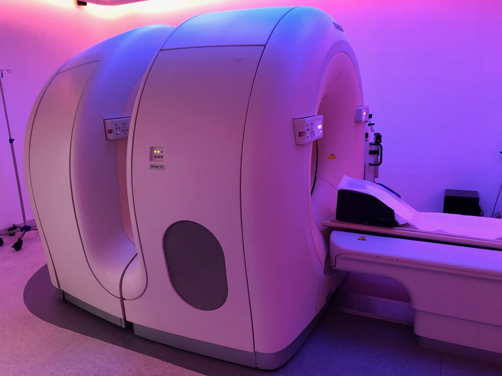
Crucially, these two gamma rays fly off in almost exactly opposite directions. The PET scanner is a large ring packed with detectors that surround the patient. When two detectors on opposite sides of the ring register a hit at the same instant, the machine knows an annihilation event occurred somewhere along the line connecting them.
By detecting millions of these paired gamma ray events, a powerful computer can reconstruct a detailed, three-dimensional image. The resulting map shows where the radiotracer has accumulated most, pinpointing the areas of high metabolic activity.

This ability to visualize function makes PET an invaluable tool in medicine, particularly for finding, staging, and monitoring cancer.
What primary aspect of tissues and organs does a PET scan visualize?
Why are PET scans particularly effective for detecting cancerous tumors?
Now that you've learned the fundamentals, you're ready to explore how this technology is applied more specifically.