Pediatric Hip Anatomy Essentials
Hip Joint Development
From Blueprint to Bone
The hip joint begins its journey early in embryonic life as a simple collection of mesenchymal cells, a type of undifferentiated tissue. Around the sixth week of gestation, this cluster of cells starts to form a cartilage model of the future hip. This model defines the shape of the joint, including the ball (femoral head) and the socket (acetabulum).
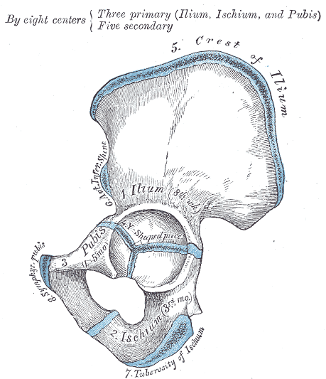
The acetabulum isn't a single bone initially. It's formed by the meeting of three separate bones: the ilium, ischium, and pubis. These three bones start as cartilage and later fuse together, creating the deep, stable socket that is a hallmark of the human hip.
The First Bony Landmarks
The process of turning cartilage into bone is called ossification. It starts at specific locations called primary ossification centers, which appear long before birth. The hip's components each have their own timeline.
The shaft of the femur, the long thigh bone, is one of the very first bones in the body to begin ossifying, starting around the seventh week of gestation. The ilium follows soon after, beginning to turn to bone in the ninth week. The ischium and pubis lag behind, starting their ossification process in the fourth and fifth fetal months, respectively.
| Bone | Primary Ossification Center Appears |
|---|---|
| Femoral Shaft | ~7th week of gestation |
| Ilium | ~9th week of gestation |
| Ischium | ~4th month of gestation |
| Pubis | ~5th month of gestation |
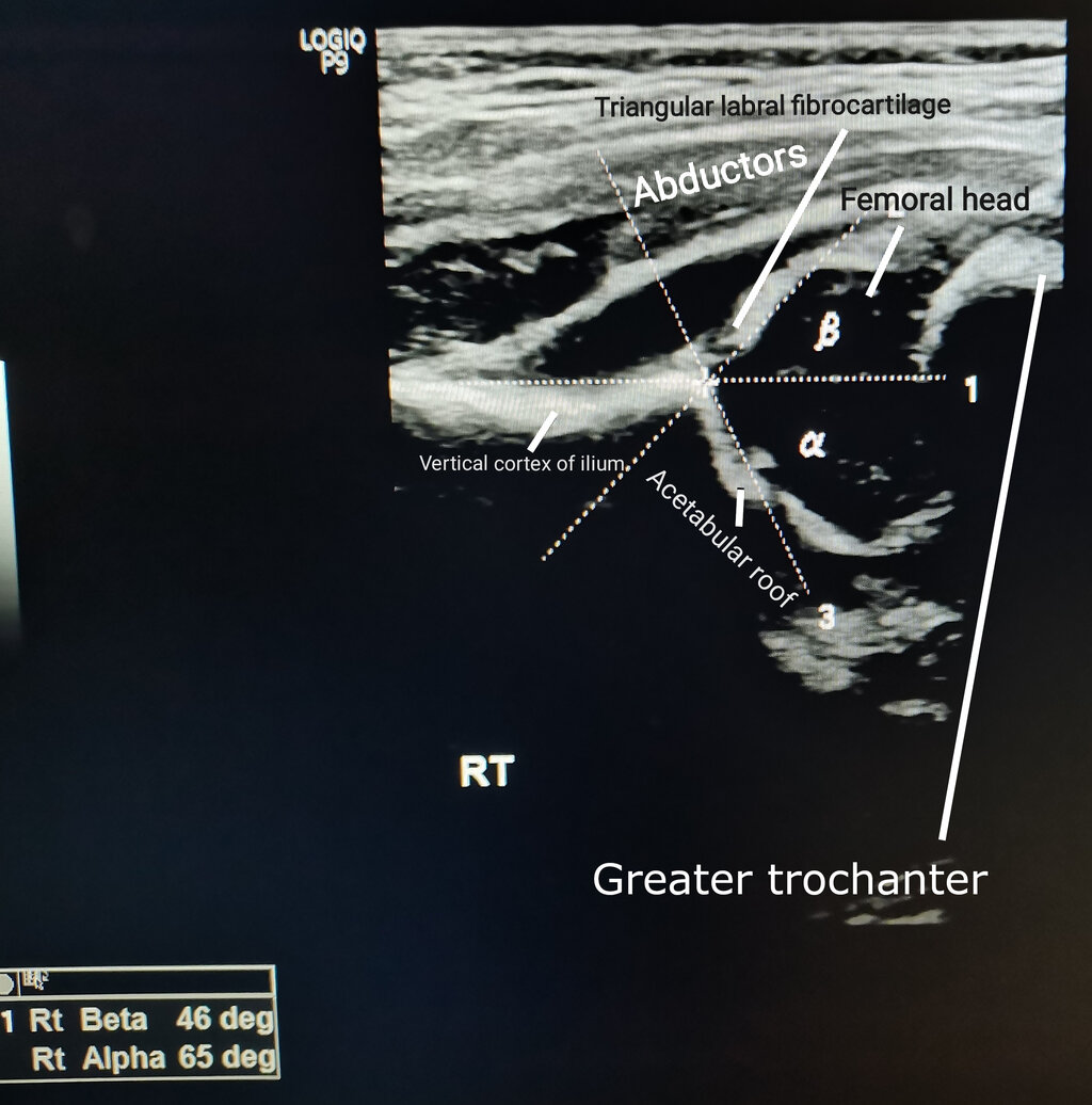
At birth, the ends of the femur and large parts of the pelvis are still cartilage. This is why a newborn's skeleton is so flexible and why it's difficult to see the full hip joint on a standard X-ray. Doctors often use ultrasound to visualize the hip in infants.
Postnatal Growth
After birth, the process of bone formation continues at secondary ossification centers. These are crucial for the growth and final shaping of the bones.
The most important secondary center for the hip is the proximal femoral epiphysis, which will become the femoral head. This center doesn't even appear until a baby is between 2 and 8 months old. Its appearance is a key milestone in hip development.
The delayed appearance of the femoral head's ossification center is normal. Its absence on an X-ray in a newborn is expected.
Other secondary centers appear later. The greater trochanter, the prominent bump on the outside of the upper femur, begins to ossify around age 4. The lesser trochanter, a smaller bump on the inside, appears much later, around age 11 to 12. These sites are important attachment points for major muscles that move the hip.
The separate bones of the acetabulum, connected by a Y-shaped piece of cartilage called the triradiate cartilage, grow throughout childhood. This cartilage acts as a growth plate for the socket itself, allowing it to enlarge to accommodate the growing femoral head. The triradiate cartilage typically closes, meaning the ilium, ischium, and pubis fuse into a single hip bone, between ages 14 and 16.
The final fusion of the femoral growth plates occurs later in adolescence. The lesser trochanter fuses to the femoral shaft around age 16-17, while the greater trochanter and the femoral head fuse around age 18. This signals the end of growth in length for the femur.
Understanding this complex timeline of bone formation and fusion is essential for pediatricians and orthopedic specialists. It allows them to read an X-ray and determine if a child's hips are developing normally or if there's a problem that needs attention.
What three bones meet and fuse to form the acetabulum, the socket of the hip joint?
Which part of the developing hip is the first to begin ossification (turning from cartilage to bone)?