Optometry Essentials
Eye Anatomy
The Eye's Window
The journey of light into the eye begins at the cornea. This transparent, dome-shaped layer on the very front of your eye acts as a clear window. But it's more than just a protective shield; it's also a powerful lens. The cornea is responsible for about two-thirds of the eye's total focusing power, bending incoming light rays to help create a sharp image.
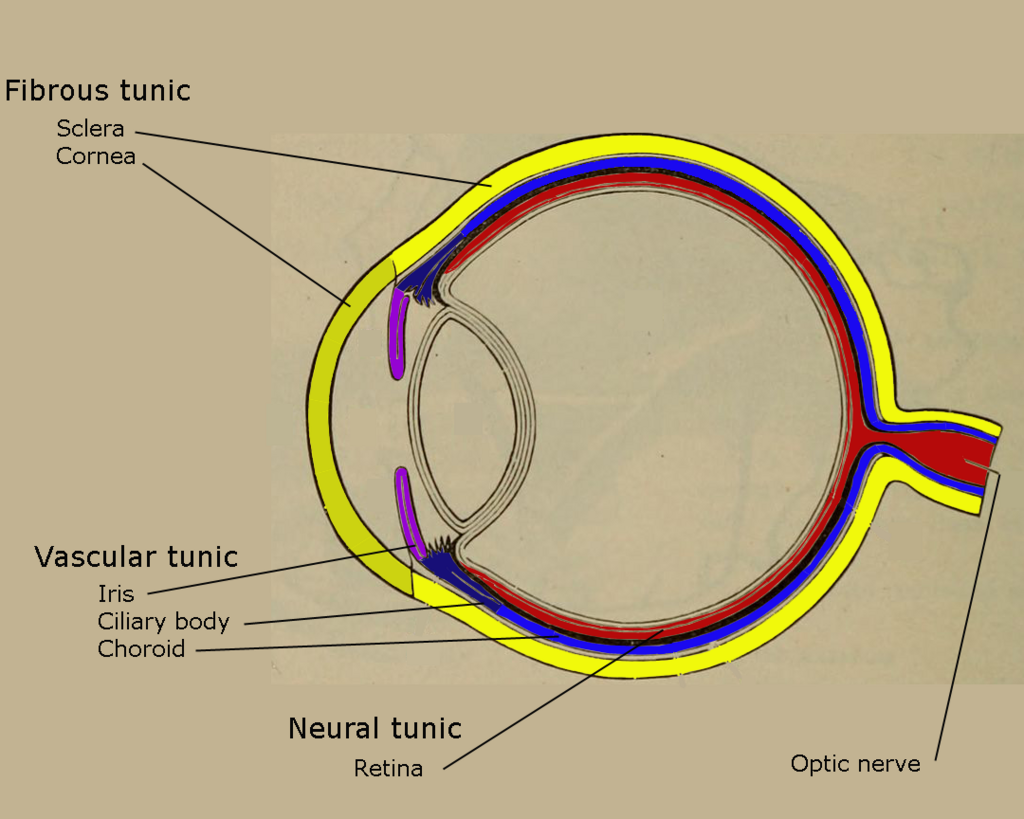
One of the most remarkable things about the cornea is that it contains no blood vessels. This is crucial for its transparency. If it had blood vessels, our vision would be obscured. Instead, it gets nutrients from the tears on its outer surface and a fluid called the aqueous humor from within the eye. This unique structure allows it to heal from minor scratches quickly without scarring.
The Adjustable Lens
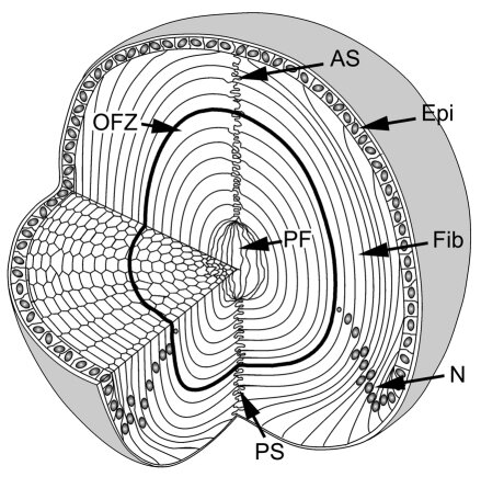
Once light passes through the cornea and the pupil, it reaches the lens. Located just behind the iris, the lens is a clear, flexible structure that fine-tunes the eye's focus. Think of it like the autofocus on a camera. It changes shape to adjust for objects at different distances.
The lens works together with the cornea to focus light correctly on the retina.
This shape-shifting is controlled by tiny ciliary muscles. When you look at something far away, the muscles relax, and the lens becomes flatter. When you focus on a nearby object, like a book, the muscles contract, causing the lens to become thicker and more rounded. This process, called accommodation, allows you to see clearly at all distances.
The Sensory Screen
At the very back of the eye is the retina, a thin layer of tissue that acts like the sensor in a digital camera. This is where light is converted into electrical signals that the brain can understand. The focused light from the cornea and lens forms an image on the retina.
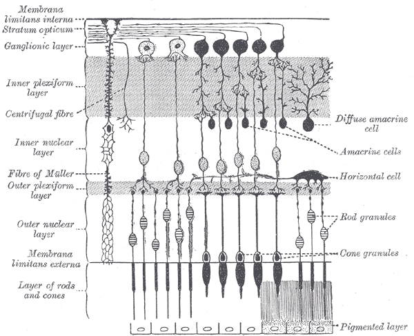
The retina is packed with millions of light-sensitive cells called photoreceptors. There are two main types:
- Rods: These are highly sensitive to light and are responsible for our vision in dim conditions and for detecting motion in our peripheral vision. They don't detect color.
- Cones: These cells work best in bright light and are responsible for sharp, detailed central vision and color perception. There are three types of cones, each sensitive to different wavelengths of light (red, green, and blue).
These photoreceptors pass their signals to other nerve cells within the retina, which begin processing the visual information before it even leaves the eye.
The Supporting Cast
Several external structures work to protect and maintain the eye. The eyelids are crucial for shielding the eye from debris and bright light. Blinking spreads tears across the eye's surface, keeping the cornea moist and clean. Eyelashes act as a first line of defense, catching dust and other particles before they can enter the eye.
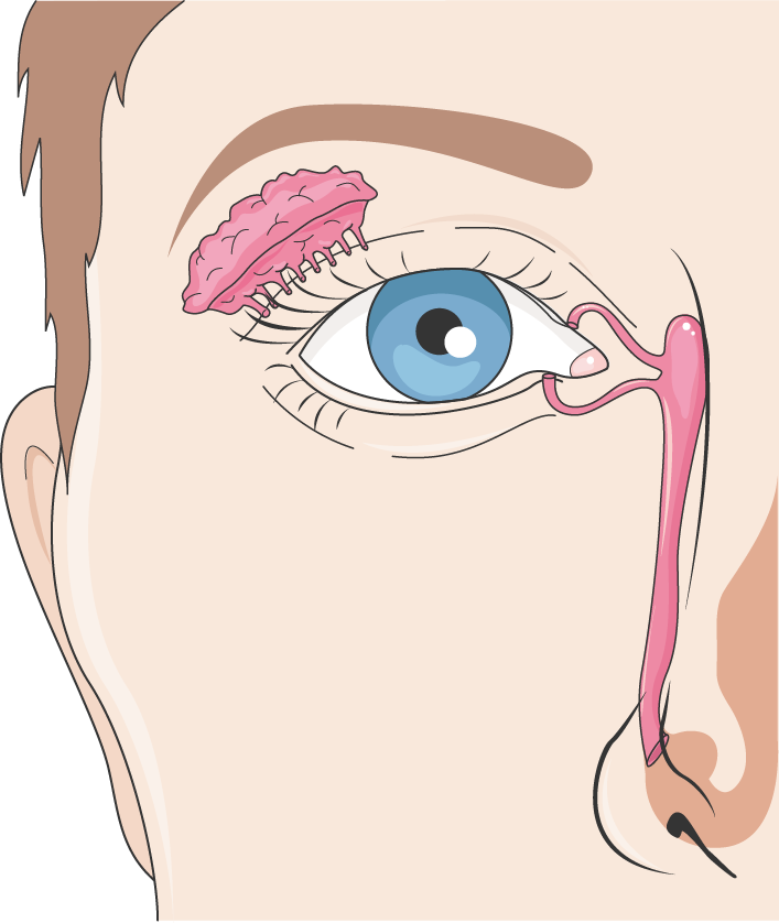
Tears are produced by the lacrimal glands, located above the outer corner of each eye. This tear film is a complex mixture that not only lubricates the eye but also provides oxygen and nutrients to the cornea and contains enzymes that protect against infection. Excess tears drain away through small ducts in the inner corner of the eyelids.
The Brain's Connection
After the retina processes the light into electrical signals, they need to get to the brain for final interpretation. This is the job of the optic nerve. It's a bundle of more than a million nerve fibers that acts as a high-speed data cable, transmitting visual information from the retina to the brain's visual cortex.
The point where the optic nerve leaves the eye creates a blind spot in our vision because there are no photoreceptors in that area.
The optic nerves from both eyes meet at a junction called the optic chiasm. Here, some of the nerve fibers cross over, so information from the right side of the visual field from both eyes is sent to the left side of the brain, and information from the left visual field goes to the right side of the brain. This complex pathway allows for depth perception and a complete, unified visual experience.
Ready to see how well you've absorbed these details? Let's check your knowledge.
What is the primary role of the cornea?
When you focus on a nearby object, the ciliary muscles contract, causing the lens to become flatter.
Each part of the eye, from the clear cornea to the complex retina, plays a vital role in the incredible process of sight.