Optimizing IGF-1 for Longevity and Performance
Cellular IGF-1 Mechanics
The Growth Signal
When you lift a heavy weight, you’re not just moving metal; you’re sending a direct chemical message to your muscle cells. One of the most important messengers in this conversation is Insulin-like Growth Factor 1, or IGF-1. This potent hormone is a primary driver of muscle hypertrophy—the process of making muscle cells larger and stronger.
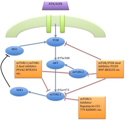
IGF-1 initiates this growth cascade by binding to its specific receptor on the surface of a muscle cell. Think of this as a key fitting into a lock. This binding event triggers a chain reaction inside the cell, a signaling pathway known as the PI3K/Akt/mTOR pathway. It’s a series of molecular dominoes, where each protein activates the next in line.
First, the activated IGF-1 receptor switches on a molecule called PI3K (Phosphoinositide 3-kinase). PI3K then activates another key protein, Akt, which is a central hub in this network. Once active, Akt performs several crucial jobs, but its most famous role in muscle growth is activating the mechanistic Target of Rapamycin complex 1, or mTORC1.
mTORC1 is the master regulator of muscle protein synthesis. When it gets the green light from Akt, it ramps up the production of new proteins, providing the raw materials to repair and build bigger, more resilient muscle fibers.
While this pathway is fundamental, where the IGF-1 signal comes from is just as important. IGF-1 operates in two distinct modes: endocrine and autocrine/paracrine.
Endocrine IGF-1 is the systemic version, produced mainly by the liver in response to growth hormone from the pituitary gland. It circulates throughout your body, promoting general growth and cellular repair. It’s what we typically measure in a blood test.
However, the real magic for muscle hypertrophy happens locally. IGF-1 is produced directly within the muscle tissue itself. This localized production is triggered not by systemic hormones, but by the physical stress of muscle contraction—a process known as mechanotransduction.
How Lifting Speaks to Your Cells
When you perform resistance exercise, the mechanical tension and metabolic stress on your muscle fibers act as a powerful local stimulus. This stress prompts the muscle cells to produce and release their own special variant of IGF-1, called Mechano-Growth Factor (MGF).
MGF is an isoform, or splice variant, of the IGF-1 gene. It’s essentially a specialized tool for the job of muscle repair and growth. Its primary role is to kickstart the muscle's repair process by activating satellite cells—the stem cells of muscle tissue. These satellite cells then fuse with existing muscle fibers, donating their nuclei and helping the fiber grow larger and stronger.
This local MGF signal is immediate and targeted, happening right where the work was done. It doesn't rely on the slower, less specific signal from the liver. This explains why targeted resistance training builds specific muscles, rather than causing uniform growth across the entire body. The growth signal is delivered directly to the tissues that earned it.
The Growth vs. Cleanup Crew
The IGF-1/Akt pathway doesn't just turn on growth; it simultaneously turns off processes that break down muscle. One of the key players in this balancing act is a group of proteins called FOXO, particularly FOXO1.
Under resting conditions, FOXO proteins reside in the cell's nucleus, where they act as transcription factors. They switch on genes associated with muscle atrophy (breakdown) and autophagy—the cellular process of cleaning up and recycling damaged components. While autophagy is essential for long-term health, having it active during a growth phase is counterproductive.
This is where Akt steps in. When Akt is activated by IGF-1, it phosphorylates FOXO1. This chemical tag acts like an eviction notice, causing FOXO1 to be kicked out of the nucleus and into the cytoplasm, where it can no longer activate its target genes. This process, called nuclear efflux, effectively slams the brakes on muscle breakdown and cleanup, allowing the mTOR-driven growth signal to dominate.
This creates a fundamental trade-off. Maximizing muscle protein synthesis via mTOR often means suppressing autophagy via FOXO inhibition. For athletes, especially during perimenopause when recovery is paramount, understanding how to cycle between these states—anabolic growth and catabolic cleanup—is key to balancing performance with long-term cellular health.
In this review, we summarized the insulin-like growth factor 1 (IGF-1)/Akt-independent activation of mammalian target of rapamycin (mTOR) signaling in muscle hypertrophy and the involvement of mTOR signaling in age-related loss of skeletal muscle function and mass and in sarcopenia.
Finally, it's important to note the interplay with insulin. Insulin and IGF-1 receptors are very similar in structure and can sometimes cross-react. High levels of insulin can weakly activate the IGF-1 receptor, and vice-versa. This molecular crosstalk is one reason why managing insulin sensitivity is so crucial for optimizing body composition and muscle health. A well-regulated system ensures that each signal is heard clearly, promoting growth when intended and allowing for repair and maintenance at other times.
What is the primary signaling pathway that Insulin-like Growth Factor 1 (IGF-1) activates to initiate muscle hypertrophy?
A weightlifter consistently trains only their biceps. Why does most of the muscle growth occur specifically in their biceps rather than uniformly throughout their body?
Understanding these cellular signals helps demystify muscle growth. It’s not just about lifting weights, but about providing the right stimulus to initiate a precise and powerful biochemical cascade.