Nursing TEAS Mastery and Exam Practice
Anatomy and Physiology Mastery
The Heart’s Two Journeys
The cardiovascular system is more than just a single pump. It's a sophisticated dual-circuit system. Every drop of blood in your body takes two distinct paths: the pulmonary circuit to the lungs and the systemic circuit to the rest of the body.
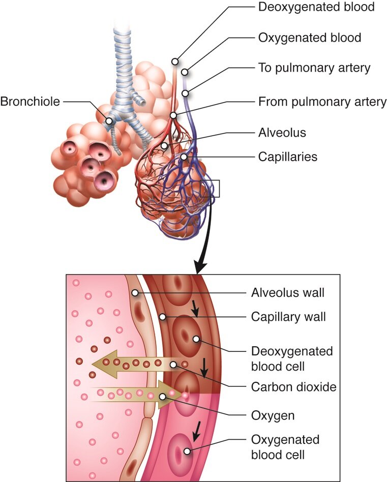
First, deoxygenated blood returns from the body and enters the right atrium. It's then pumped by the right ventricle to the lungs via the pulmonary artery. This is the only artery in the body that carries deoxygenated blood. In the lungs, the blood offloads carbon dioxide and picks up fresh oxygen. Now oxygenated, it returns to the left atrium through the pulmonary veins.
This oxygen-rich blood then begins its journey through the systemic circuit. The left ventricle, the heart's most powerful chamber, pumps this blood into the aorta, the body's largest artery. From there, it travels to every organ and tissue, delivering oxygen and nutrients. After this exchange, the now deoxygenated blood makes its way back to the right atrium, and the cycle begins again.
This entire process is driven by a precise electrical conduction system. The heartbeat originates in the , the heart's natural pacemaker. The signal spreads across the atria, causing them to contract, then travels to the atrioventricular (AV) node. After a brief delay, the impulse moves down to the ventricles, causing them to contract and pump blood out to the lungs and body. An electrocardiogram (EKG or ECG) is simply a recording of this electrical activity.
The Body's pH Balance
Your lungs do more than just supply oxygen. They play a critical role in maintaining the body's acid-base balance by regulating carbon dioxide levels. This process is crucial for keeping your blood pH within a very narrow, stable range of 7.35 to 7.45.
When CO2 mixes with water in the blood, it forms carbonic acid (H₂CO₃), a weak acid. This acid then dissociates into hydrogen ions (H⁺) and bicarbonate ions (HCO₃⁻). The concentration of hydrogen ions is what determines the blood's pH. More H⁺ means lower pH (more acidic), and less H⁺ means higher pH (more alkaline).
The relationship is governed by the bicarbonate buffer system, a rapid and elegant chemical equilibrium.
If your blood becomes too acidic (acidosis), your brain signals your respiratory muscles to breathe faster and deeper. This process, called hyperventilation, expels more CO2. According to the equation, reducing CO2 shifts the equilibrium to the left, which consumes H⁺ ions and raises the blood pH back to normal.
Conversely, if your blood is too alkaline (alkalosis), your breathing slows down. This hypoventilation allows CO2 to accumulate, shifting the equilibrium to the right, which increases the concentration of H⁺ ions and lowers the pH.
Chemical Digestion and Absorption
The gastrointestinal system is a disassembly line. It breaks down large food molecules into smaller units that can be absorbed into the bloodstream. While mechanical digestion (chewing, churning) starts the process, chemical digestion does the fine-detail work using specialized enzymes.
It begins in the mouth with salivary amylase breaking down starches. In the stomach, pepsin, activated by hydrochloric acid, starts protein digestion. The real powerhouse of chemical digestion, however, is the small intestine. Here, enzymes from the pancreas and the intestinal wall break down carbohydrates, proteins, and fats into their simplest forms.
| Enzyme | Source | Macronutrient Digested |
|---|---|---|
| Amylase | Salivary Glands, Pancreas | Carbohydrates |
| Pepsin | Stomach | Proteins |
| Trypsin | Pancreas | Proteins |
| Lipase | Pancreas | Fats (Lipids) |
| Lactase/Sucrase | Small Intestine | Carbohydrates (Sugars) |
Once digestion is complete, absorption occurs. The vast majority of nutrients are absorbed in the small intestine, whose inner surface is covered with millions of tiny, finger-like projections called . These structures dramatically increase the surface area available for absorption. Monosaccharides (from carbs), amino acids (from proteins), fatty acids (from fats), vitamins, minerals, and water all pass through the cells of the villi and enter the bloodstream or lymphatic system to be transported throughout the body.
The Spark of Movement
Every voluntary movement you make, from blinking to sprinting, begins with a signal from your brain. This signal travels down a motor neuron to the muscle fiber it controls. The point of contact between the nerve and the muscle is called the .
When the nerve impulse reaches the end of the neuron, it triggers the release of a neurotransmitter called acetylcholine (ACh). ACh crosses the tiny gap (the synapse) and binds to receptors on the muscle fiber. This binding action generates an electrical signal in the muscle cell, which is the direct trigger for contraction.
Once triggered, the muscle contracts via the sliding filament theory. Inside each muscle fiber are long protein filaments called actin (thin filaments) and myosin (thick filaments), arranged in repeating units called sarcomeres. The electrical signal causes calcium ions to be released within the muscle cell. This calcium allows the myosin heads to grab onto the actin filaments, forming cross-bridges. The myosin heads then pull the actin filaments inward, causing the sarcomere to shorten. As millions of sarcomeres shorten in unison, the entire muscle contracts.
What is the primary function of the villi and microvilli in the small intestine?
Which of the following correctly traces the path of oxygenated blood?