New Treatments for Glioblastoma
Introduction to Glioblastoma Multiforme
What is Glioblastoma?
Glioblastoma, often called glioblastoma multiforme (GBM), is the most common and most aggressive type of cancerous tumor that begins in the brain. It's known as a primary brain tumor, which means it originates in the brain itself, rather than spreading there from another part of the body.
Glioblastoma
noun
An aggressive type of cancer that can occur in the brain or spinal cord. Glioblastoma forms from cells called astrocytes that support nerve cells.
These tumors are notorious for their rapid growth and their tendency to invade nearby brain tissue. They are made up of a mix of different cell types, which makes them particularly challenging to understand and treat.
Who Gets Glioblastoma?
While GBM can affect people of any age, it is most commonly diagnosed in older adults, typically between the ages of 65 and 75. It is slightly more common in men than in women.
Overall, glioblastoma is a rare disease, affecting about 3 out of every 100,000 people. However, despite its rarity, it accounts for nearly half of all primary malignant brain tumors.
For the vast majority of cases, the cause of GBM is unknown. There are very few established risk factors. The most significant one is prior exposure to high doses of ionizing radiation to the head, often from treatment for a previous cancer. Age is the other main risk factor, with the likelihood of developing GBM increasing as we get older.
Inside the Tumor
One of the defining features of glioblastoma is its diffuse nature. The tumor doesn't grow as a neat, contained ball. Instead, it has finger-like tentacles that infiltrate deep into the surrounding brain tissue. This makes it impossible to remove completely through surgery alone, as cancerous cells are always left behind.
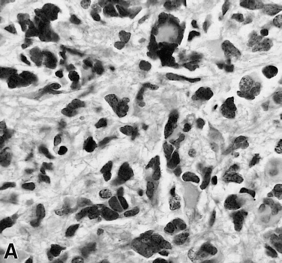
Another challenge is the tumor's heterogeneity. This means the cells within a single glioblastoma are not all the same. They can have different genetic mutations, making some cells resistant to certain therapies while others are not. This diversity allows the tumor to adapt and continue growing even when under attack.
At the genetic level, glioblastomas are complex. They often involve mutations in several key genes that normally control how cells grow and divide. Common alterations are found in genes like:
- EGFR (Epidermal Growth Factor Receptor): This gene helps cells grow. In many GBMs, it's amplified, meaning there are too many copies of it, leading to out-of-control growth.
- TP53: This is a tumor suppressor gene, often called the "guardian of the genome." When it's mutated, it can no longer stop cells from becoming cancerous.
- PTEN: Another tumor suppressor gene that, when mutated, fails to put the brakes on cell division.
Symptoms and Diagnosis
The symptoms of glioblastoma depend heavily on the tumor's size and location in the brain. As the tumor grows, it increases pressure inside the skull, which can lead to general symptoms like persistent headaches, nausea, and vomiting.
More specific symptoms relate to which part of the brain is affected. For example:
- A tumor in the frontal lobe might cause personality changes, mood swings, or muscle weakness.
- A tumor in the temporal lobe could lead to problems with speech or memory.
- A tumor in the parietal lobe may affect sensation, like touch or pressure.
Seizures are also a common presenting symptom, occurring when the tumor's abnormal electrical activity disrupts normal brain function.
Diagnosis usually begins when a person experiences neurological symptoms that prompt a doctor to order a brain scan.
The primary tools for diagnosis are imaging tests. Magnetic Resonance Imaging (MRI) is the preferred method because it provides detailed pictures of the brain's soft tissues. On an MRI, a glioblastoma often appears as a "ring-enhancing" lesion—a bright ring of contrast agent dye surrounding a darker, necrotic (dead tissue) core.
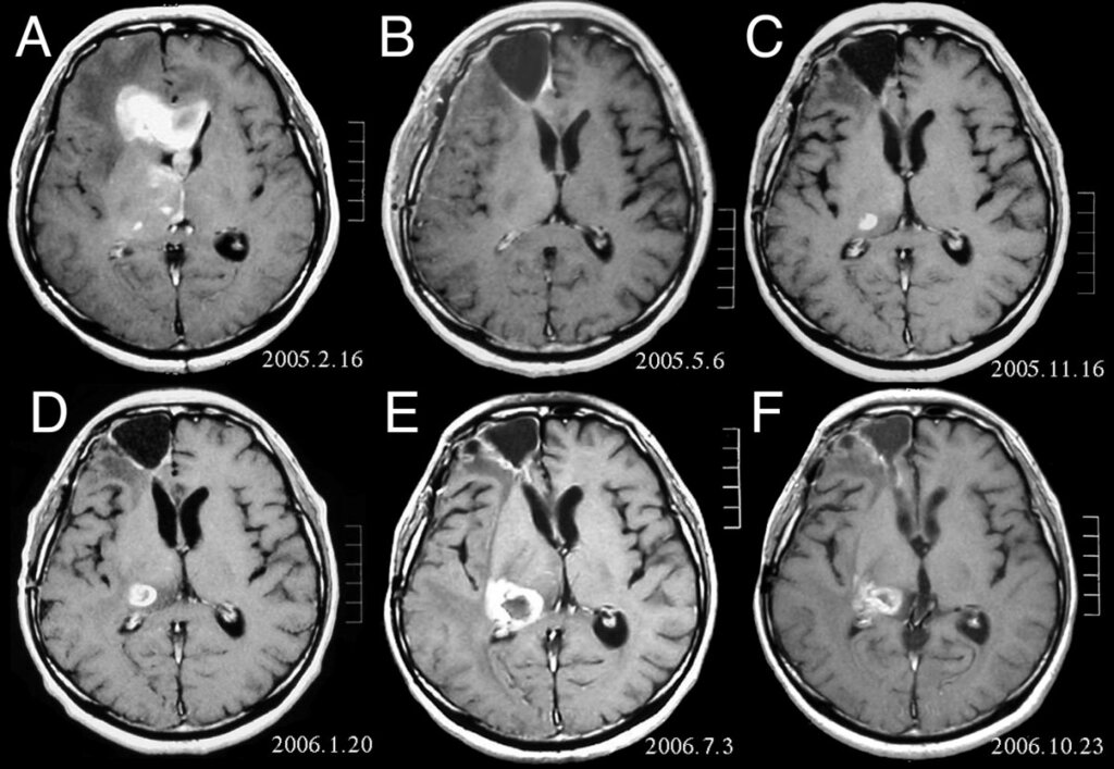
While an MRI can strongly suggest a glioblastoma, a definitive diagnosis requires a biopsy. This involves surgically removing a small sample of the tumor tissue, which is then examined under a microscope by a pathologist. The pathologist looks for characteristic features of GBM cells to confirm the diagnosis.