Medical Imaging Showdown MRI CT X-ray
X-ray Imaging
Seeing with Shadows
X-ray imaging is like creating a shadow picture of the inside of your body. It uses a special kind of high-energy light, called X-rays, that we can't see with our eyes. This light can easily pass through soft tissues like skin and muscle, but it gets stopped by denser materials, especially bone and metal.
When you get an X-ray, a machine sends a quick burst of these rays through a part of your body. On the other side, a detector (which used to be special film, but is now usually digital) captures the rays that make it through. Bones show up as bright white areas because they block most of the X-rays. Softer tissues appear in shades of gray, and air-filled spaces, like your lungs, look black because almost all the X-rays pass through them.
This simple process gives doctors a clear, two-dimensional picture of your bones and certain other structures, making it a powerful tool for diagnosis.
Creating the Rays
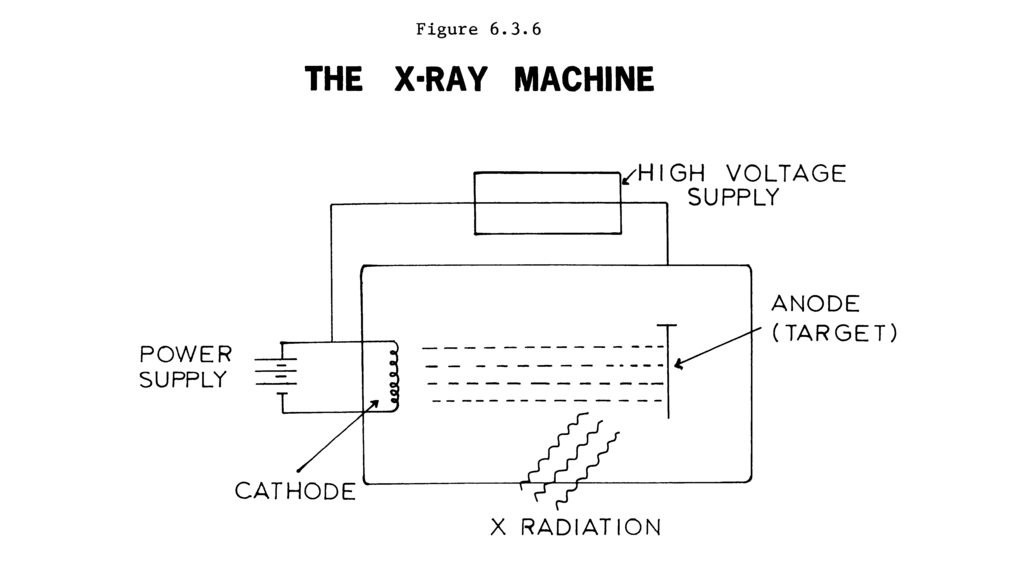
The magic happens inside a device called an X-ray tube. It’s a vacuum-sealed tube with two key components: a negative electrode (cathode) and a positive electrode (anode). When the machine is turned on, the cathode heats up and releases a stream of electrons. A high voltage then pulls these electrons across the vacuum at incredible speeds until they smash into the anode, which is a metal target.
This high-energy collision is what generates the X-rays. The sudden stop converts the electrons' kinetic energy into a tiny amount of X-ray energy and a lot of heat. The X-rays are then focused into a beam and directed toward the patient.
Common Clinical Uses
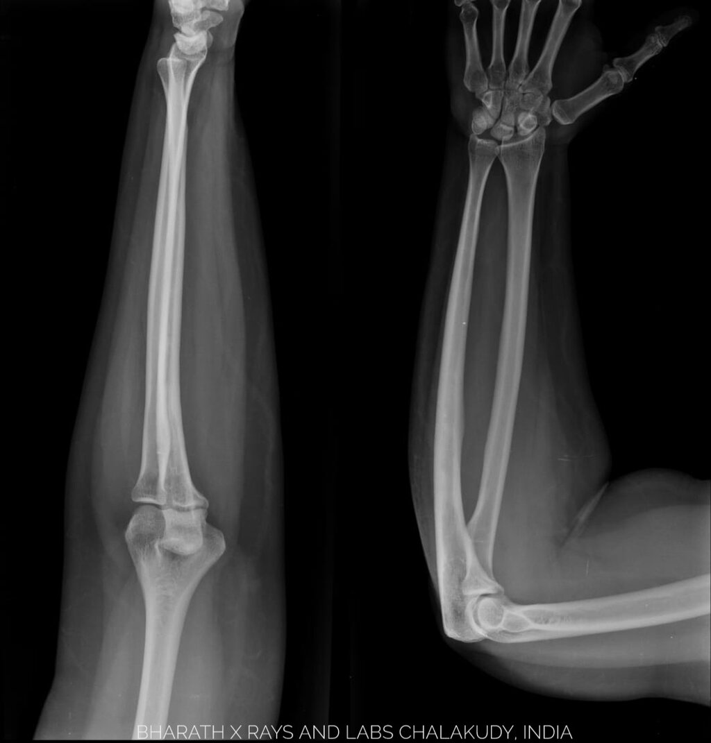
You've most likely encountered X-rays for checking broken bones. Their ability to show bone detail with high clarity makes them the go-to method for diagnosing fractures. A quick look at an X-ray can tell a doctor the exact location and severity of a break.
But X-rays aren't just for bones. Chest X-rays are one of the most common medical imaging tests performed. They can help detect lung diseases like pneumonia, which shows up as a whitish area in the normally dark lung fields. They can also reveal an enlarged heart or other issues within the chest cavity.
Strengths and Weaknesses
X-ray imaging remains a cornerstone of medicine for several key reasons. But it also has important limitations to consider.
| Advantages | Limitations |
|---|---|
| Fast: The process takes only a few minutes. | Low Soft Tissue Contrast: Struggles to show detail in muscles, ligaments, and organs. |
| Inexpensive: Costs less than other advanced imaging techniques. | Radiation Exposure: Uses ionizing radiation, which carries a small risk. |
| Widely Available: Most clinics and hospitals have X-ray equipment. | 2D Images: Flattens 3D structures into one plane, which can sometimes hide problems. |
The use of ionizing radiation is the most significant limitation. While the dose from a single X-ray is very low and the benefit almost always outweighs the risk, doctors are careful to only order them when necessary. This is especially true for children and pregnant women. However, for a quick and effective look at the body's densest structures, X-ray imaging is still an essential and invaluable tool.