Mechanics of the Human Heart
Cardiac Electrical Conduction
The Heart's Internal Pacemaker
The heart's beat isn't commanded by the brain; it generates its own rhythm. This intrinsic electrical system ensures your heart contracts in a coordinated, efficient sequence, pushing blood throughout your body. The entire process starts in a small cluster of specialized cells in the right atrium called the .
The conduction system within the heart is vital because it permits a rapid and organized depolarization of ventricular myocytes that is necessary for the efficient generation of pressure during systole.
These pacemaker cells in the SA node have a different kind of action potential compared to the contractile muscle cells (myocytes) that make up most of the heart. Instead of a stable resting phase, they undergo a slow, spontaneous depolarization. Once they reach a threshold, they fire, creating an electrical wave that spreads across both atria, causing them to contract and push blood into the ventricles.
As the wave of depolarization from the SA node reaches the floor of the right atrium, it encounters the Atrioventricular (AV) node. Here, the signal encounters a crucial, intentional traffic jam.
The AV node delays the electrical impulse by about 120 milliseconds. This pause is vital; it gives the atria enough time to finish contracting and completely fill the ventricles with blood before they are stimulated to contract.
The Ventricular Superhighway
After the delay, the AV node passes the impulse to a high-speed distribution network. The signal travels down the (pronounced 'hiss'), which runs through the septum separating the ventricles. This bundle then splits into right and left bundle branches, which carry the impulse toward the bottom (apex) of the heart.
The bundle branches terminate in a vast network of fibers that spread throughout the ventricular walls, called the . These fibers conduct the electrical impulse extremely rapidly, about four times faster than any other tissue in the heart. This speed ensures that all the contractile cells in the ventricles depolarize and contract almost simultaneously, from the bottom up. This coordinated, powerful squeeze is what efficiently ejects blood into the aorta and pulmonary artery.
This bottom-up contraction is like squeezing a tube of toothpaste from the bottom, ensuring maximum ejection of its contents.
Reading the Signals
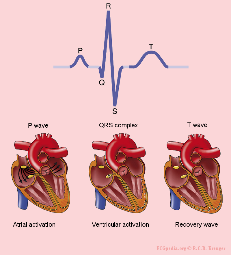
An electrocardiogram (ECG or EKG) is a recording of this electrical activity. Each part of the ECG waveform corresponds to a specific event in the conduction cycle.
| Waveform | Electrical Event | Mechanical Event |
|---|---|---|
| P Wave | Atrial depolarization (signal from SA node spreads) | Atrial contraction |
| QRS Complex | Ventricular depolarization (signal spreads through Purkinje fibers) | Ventricular contraction (systole) |
| T Wave | Ventricular repolarization (ventricles reset electrically) | Ventricular relaxation (diastole) |
By analysing the shape and timing of these waves, doctors can diagnose a wide range of cardiac issues, from arrhythmias to damage from a heart attack. The simple squiggles on an ECG provide a direct window into the elegant electrical symphony that keeps us alive.
Ready to test your knowledge of the heart's electrical system?
Where does the electrical impulse that triggers a normal heartbeat originate?
What is the primary function of the Atrioventricular (AV) node in the heart's conduction system?
Understanding this pathway is fundamental to cardiology, as disruptions in this sequence are the root cause of many heart rhythm disorders.