Mechanics and Regulation of the Human Heartbeat
Electrical Conduction Pathways
The Heart's Pacemaker
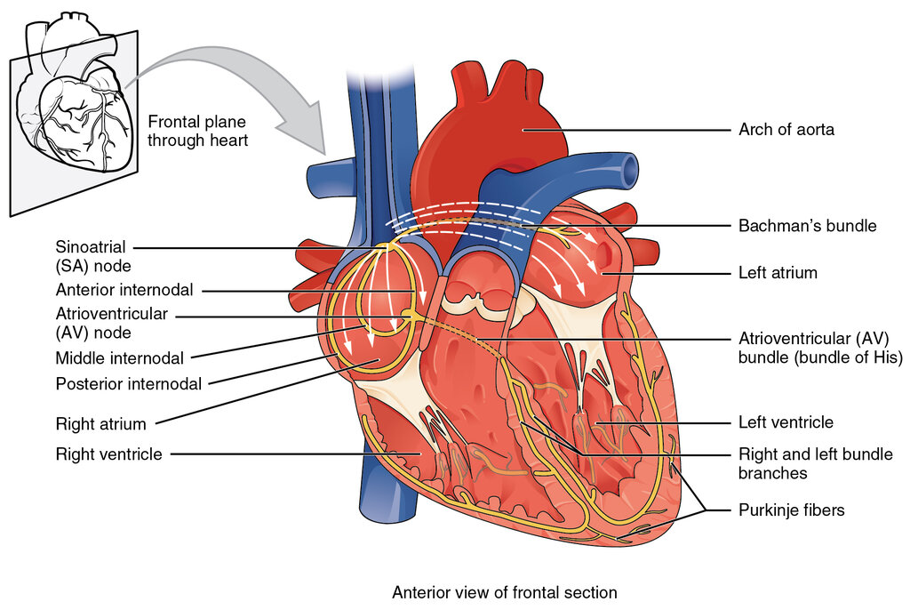
Every heartbeat begins as an electrical spark. This spark doesn't come from your brain or spinal cord; it originates within the heart muscle itself, in a tiny cluster of specialized cells called the sinoatrial (SA) node. Located in the upper wall of the right atrium, the is the heart's natural pacemaker.
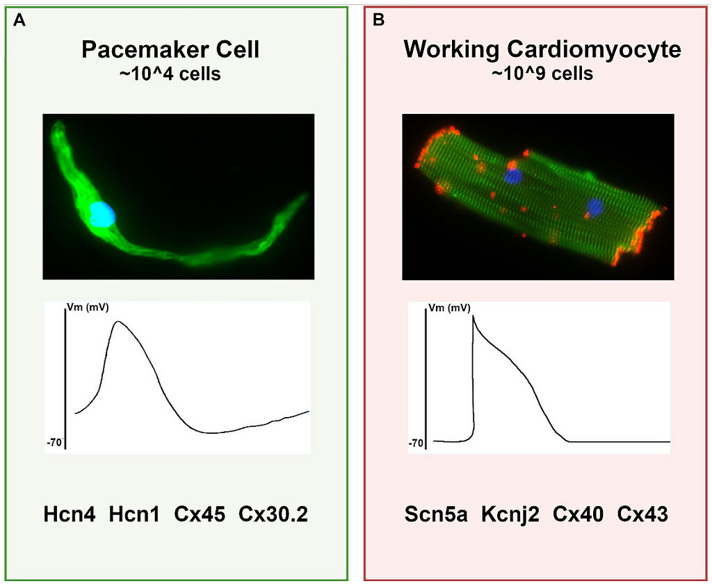
Unlike regular muscle cells that need a nerve signal to contract, pacemaker cells in the SA node spontaneously depolarize. This process, called the pacemaker potential, isn't driven by the fast-acting sodium channels found elsewhere. Instead, it relies on a slow, steady leak of positive ions, primarily through channels humorously called 'funny channels', followed by an influx of calcium ions (). This calcium influx is what triggers the action potential, creating the electrical impulse.
The SA node fires at the fastest intrinsic rate in the heart, typically 60 to 100 times per minute. This inherent speed allows it to control the other potential pacemaker sites in the heart through a process called s. Essentially, the SA node's rapid firing resets the slower-firing cells before they have a chance to generate their own beat, ensuring a single, coordinated rhythm.
The Crucial Delay
Once the SA node fires, the electrical wave spreads across the atria, causing them to contract and push blood into the ventricles. The signal then converges on another key structure: the atrioventricular (AV) node, located near the center of the heart between the atria and ventricles.
The electrical stimulus from the SA node eventually reaches the AV node and is delayed briefly so that the contracting atria have enough time to pump all the blood into the ventricles.
The AV node's primary job is to act as a gatekeeper. It intentionally slows down the electrical signal for about 0.12 seconds. This pause, known as the s, is not a malfunction; it's a critical design feature. It gives the atria enough time to finish contracting and completely fill the ventricles before they are signaled to contract.
This delay happens because the cells in the AV node are smaller, have fewer gap junctions (the channels connecting cells), and rely on the same slow calcium currents as the SA node. This combination creates a biological bottleneck, ensuring the cardiac cycle unfolds in the correct sequence.
The Ventricular Superhighway
After its brief pause at the AV node, the impulse is ready for rapid distribution throughout the ventricles. It travels down a specialized pathway called the Bundle of His, which quickly splits into the right and left bundle branches. These branches run along the interventricular septum, the wall separating the two ventricles.
The bundle branches terminate in a vast network of fibers that spread throughout the ventricular walls, called the Purkinje fibers. These fibers are the expressways of the heart's conduction system. They are large cells with many gap junctions, allowing them to conduct the electrical impulse up to four times faster than any other cell type in the heart.
| Pathway | Conduction Velocity (m/s) |
|---|---|
| SA Node | ~0.05 |
| Atrial Muscle | ~1.0 |
| AV Node | ~0.05 |
| Bundle of His | ~1.0 |
| Purkinje Fibers | ~4.0 |
This incredible speed ensures that all parts of both ventricles contract in a synchronized, coordinated fashion. The depolarization wave starts at the interventricular septum, travels down to the apex (the bottom tip) of the heart, and then sweeps upwards along the outer walls. This bottom-up contraction is the most efficient way to eject blood from the ventricles into the aorta and pulmonary artery, much like squeezing a tube of toothpaste from the bottom.
Now, let's test your understanding of this intricate electrical pathway.
What is the primary function of the sinoatrial (SA) node?
The delay of the electrical signal at the atrioventricular (AV) node is a critical design feature, not a malfunction.