Mastering the Architecture of Life
Protein Folding Dynamics
The Folding Puzzle
A newly made polypeptide chain is a long, flexible string of amino acids. Its function, however, depends entirely on it collapsing into a very specific, stable three-dimensional shape. How does it find this one correct shape out of a seemingly infinite number of possibilities? This isn't a trivial question.
In the 1960s, a molecular biologist named Cyrus Levinthal calculated that if a small protein of 100 amino acids tried to find its final shape by randomly sampling every possible conformation, it would take longer than the age of the universe. Yet, in our cells, proteins fold in microseconds to seconds. This discrepancy is known as and it tells us something profound: protein folding is not a random search. It's a directed process, guided by the laws of thermodynamics.
The final, folded state of a protein is the one with the lowest Gibbs free energy. The process is spontaneous, driven by the desire to reach the most stable energetic state possible in its aqueous environment.
This energy landscape illustrates the folding process. The unfolded protein sits at the top of a 'funnel', with high energy and many possible shapes. As it folds, it tumbles down the funnel, losing energy and sampling fewer conformations until it settles into its stable, native state at the bottom.
Local Structures First
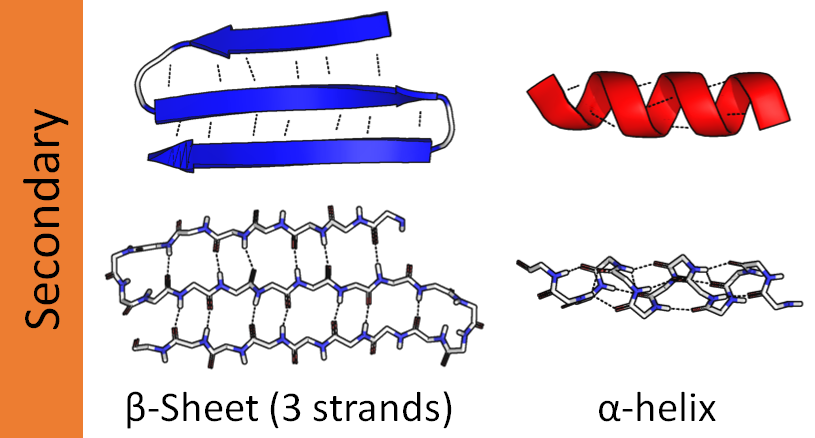
The journey down the energy funnel isn't a straight drop. It begins with the formation of local, stable structures called secondary structures. These are recurring motifs primarily stabilized by hydrogen bonds between atoms in the polypeptide backbone.
The two most common secondary structures are the α-helix and the β-sheet. An α-helix is a right-handed coil, like a spring, where hydrogen bonds form between an amino acid and another one four residues down the chain. A β-sheet is formed when different segments of the chain, called β-strands, align side-by-side. Hydrogen bonds form between these adjacent strands, creating a pleated, sheet-like structure.
The Final Fold and its Forces
Once secondary structures form, the protein collapses into its final, compact three-dimensional shape, known as the tertiary structure. This global fold is held together by a variety of interactions between the amino acid side chains (R-groups).
A primary driver of this collapse is the —the tendency of nonpolar side chains to avoid water. They bury themselves in the protein's core, leaving the polar, hydrophilic side chains on the surface to interact with the surrounding water. Other forces at play include:
- Ionic bonds: Attractions between positively and negatively charged side chains.
- Hydrogen bonds: Interactions between polar side chains.
- Van der Waals forces: Weak, transient attractions between all atoms.
- Disulfide bridges: Strong covalent bonds that can form between two cysteine residues, locking parts of the protein together.
A Helping Hand
While the folding pathway is thermodynamically favorable, the cellular environment is incredibly crowded. A newly synthesized polypeptide is at risk of sticking to other molecules and aggregating before it can fold correctly. To prevent this, cells use specialized proteins called molecular chaperones.
Chaperones don't dictate the final folded shape. They simply provide a safe environment for the polypeptide to fold, preventing aggregation and helping to untangle misfolded intermediates.
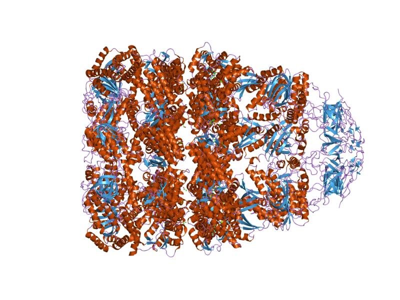
Some chaperones bind to exposed hydrophobic patches on unfolded proteins, preventing them from clumping together. A more complex class, known as chaperonins, form elaborate, barrel-shaped chambers. An unfolded protein enters the chamber, a 'lid' seals it inside, and within this isolated space, it can fold without interference. After a few seconds, the lid comes off, and the now-folded protein is released. One of the best-studied examples is the complex in bacteria.
Sometimes, despite all these systems, proteins fail to fold correctly or become unfolded due to cellular stress like heat. These misfolded proteins are often tagged for destruction, but if this quality control system fails, they can accumulate and cause serious problems.
Misfolded proteins often expose their sticky hydrophobic cores, causing them to clump together into large, insoluble aggregates. One particularly stable and dangerous form of aggregate is the , a highly ordered structure rich in β-sheets. The accumulation of these amyloid plaques is a hallmark of several neurodegenerative diseases, including Alzheimer's and Parkinson's disease, as they disrupt normal cellular function.
Now, let's test your understanding of these complex dynamics.
Levinthal's paradox highlights a significant discrepancy between the calculated time for a protein to fold by random search and the actual time it takes in a cell. What does this paradox imply about the process of protein folding?
What is the primary driving force that causes a polypeptide chain to collapse into its compact tertiary structure?
Understanding how a linear chain becomes a functional machine is central to all of biology. From the initial formation of local helices to the guiding hand of chaperones, protein folding is a rapid, specific, and essential process.