Male Reproductive Health and Anatomy
Anatomical Architecture
Inside the Sperm Factory
The testes are more than just two oval glands; they are intricate factories optimized for sperm production, or spermatogenesis. Internally, each testis is divided into about 250-300 compartments called testicular lobules. Think of them as individual production floors in the factory.
Each lobule is packed with one to four tightly coiled tubes known as seminiferous tubules. If you were to uncoil and lay out all the tubules from a single testis, they would stretch for hundreds of meters. This immense surface area is where the magic happens. The walls of these tubules are lined with a specialized epithelium containing two crucial cell types: Sertoli cells, which nourish developing sperm, and the sperm cells themselves at various stages of maturation.
Nestled between the seminiferous tubules are the interstitial cells of Leydig. These cells have a completely different job: they are the primary source of testosterone, the hormone that drives the development of male secondary sexual characteristics and is essential for spermatogenesis itself. This architectural arrangement is incredibly efficient, placing hormone production right next to the cells that need it most.
Critical Climate Control
Sperm production is a delicate process that requires a temperature about 2-4°C below core body temperature. The scrotum provides this precise environment through a combination of clever anatomical features.
First, the dartos muscle, a layer of smooth muscle in the scrotal wall, contracts in the cold, causing the skin to wrinkle. This reduces the surface area available for heat loss. Conversely, it relaxes when warm to increase surface area. Second, the cremaster muscle, an extension of the internal oblique abdominal muscle, acts like an elevator for the testes. It contracts to pull the testes closer to the body for warmth and relaxes to let them descend away from the body to cool down.
The most sophisticated mechanism, however, is the pampiniform plexus—a network of veins that wraps around the testicular artery. This arrangement acts as a countercurrent heat exchanger. Warm arterial blood flowing down to the testes transfers its heat to the cooler venous blood returning to the body. By the time the arterial blood reaches the testes, it has been pre-cooled, protecting the sensitive sperm-producing tissues.
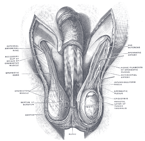
These thermoregulatory systems work in constant concert, making subtle adjustments to maintain the perfect temperature for fertility.
The Journey Outward
Once produced, sperm are not yet ready for fertilization. They must undergo a maturation process while traveling through a series of ducts. The journey begins in the epididymis, a single, tightly coiled tube about 6 meters (20 feet) long that cups the back of the testis. Sperm spend about two weeks slowly moving through the epididymis, during which they mature, gain motility, and become capable of fertilization.
The wall of the epididymis contains smooth muscle that helps propel the sperm along. Its lining is made of pseudostratified columnar epithelium with long microvilli called stereocilia. These aren't for movement; they absorb excess fluid and pass nutrients to the stored sperm.
From the epididymis, sperm enter the vas deferens (or ductus deferens). This is a much thicker, muscular tube that travels up from the scrotum, into the pelvic cavity, and loops over the bladder. The wall of the vas deferens has three layers of smooth muscle, which contract vigorously during ejaculation to propel sperm forward via peristalsis.
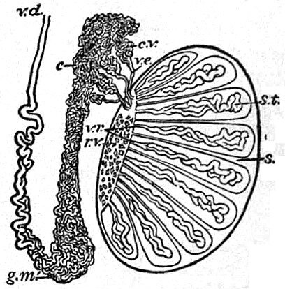
Before ejaculation, the vas deferens widens into an ampulla, where it joins with the duct from the seminal vesicle to form the ejaculatory duct. This short duct passes through the prostate gland and empties into the urethra.
The entire ductal system—from the seminiferous tubules to the urethra—is a continuous, one-way street designed to produce, mature, store, and transport sperm.
The final part of the journey is through the urethra, a tube that serves both the reproductive and urinary systems. It extends from the bladder through the prostate gland and penis to the outside of the body. During ejaculation, a sphincter at the base of the bladder contracts, preventing urine from mixing with semen.
The Chemical Cocktail Crew
Sperm themselves make up only a small fraction of the final ejaculate volume. The rest is seminal fluid, a complex mixture produced by three accessory glands: the seminal vesicles, the prostate gland, and the bulbourethral glands. Each contributes essential ingredients to the cocktail.
The seminal vesicles are a pair of glands on the posterior surface of the bladder. They secrete a thick, alkaline fluid that contains fructose (an energy source for sperm), prostaglandins (which may help sperm motility and viability), and a coagulating agent. This fluid constitutes about 70% of semen volume.
The prostate gland adds a milky, slightly acidic fluid containing citrate (another nutrient source) and several enzymes, including prostate-specific antigen (PSA), which helps liquefy the semen after ejaculation.
Finally, the tiny, pea-sized bulbourethral (Cowper's) glands, located at the base of the penis, produce a thick, clear mucus. This fluid is released prior to ejaculation to neutralize any acidic urine remaining in the urethra and to lubricate the tip of the penis.
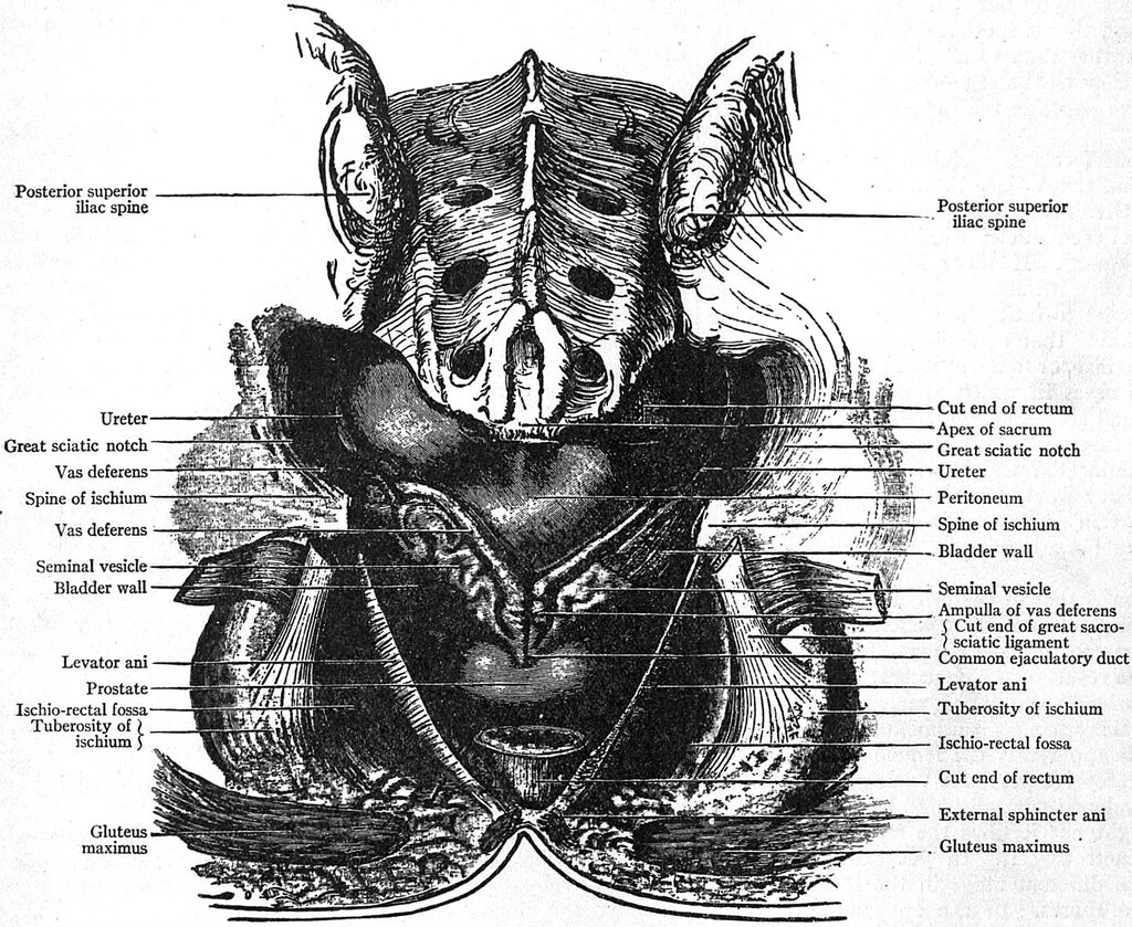
Together, these glands create a fluid medium that nourishes the sperm, protects them from the acidic environment of the male urethra and female vagina, and facilitates their movement towards the egg. The structural arrangement of these glands along the ductal path ensures their secretions are added at precisely the right moment.
Now, let's test your understanding of this intricate system.
What is the primary function of the interstitial cells of Leydig, which are found nestled between the seminiferous tubules?
Which structure acts as a countercurrent heat exchanger, cooling arterial blood before it reaches the testes?
The male reproductive system is a marvel of anatomical engineering, where each structure's form is perfectly tailored to its physiological function, from microscopic tubules to complex vascular networks.