Lymphomatoid Papulosis Management
Subtype Classification Spectrum
The LyP Subtype Spectrum
Lymphomatoid Papulosis (LyP) presents a fascinating clinical and histologic paradox: a condition that looks malignant under the microscope but typically follows a benign, chronic course. The 2018 WHO-EORTC classification expanded our understanding of LyP, providing a framework that connects its diverse histopathologic presentations to distinct clinical morphologies. This system moves beyond the classic Types A, B, and C to include newer, rarer variants, allowing for more precise diagnosis and management.
Classic Presentations: Types A, B, and C
Type A is the most common presentation, accounting for about 80% of cases. Histologically, it's defined by a wedge-shaped, polymorphous dermal infiltrate. Within this infiltrate, you'll find scattered or small clusters of large, atypical CD30+ lymphoid cells mixed with a significant population of inflammatory cells, including neutrophils, eosinophils, and histiocytes. This busy background can sometimes obscure the neoplastic cells, but it's a hallmark of the subtype. Clinically, this corresponds to the classic presentation of recurrent, self-healing papules and nodules.
Type B is the great mimicker. It resembles (MF), showing prominent epidermotropism where atypical, small- to medium-sized lymphocytes with cerebriform nuclei infiltrate the epidermis. These cells often form a band-like pattern in the papillary dermis. Unlike other LyP types, the CD30 expression in Type B can be sparse, with only a few scattered positive cells. This subtype clinically presents as scaly papules or patches, further strengthening its resemblance to patch- or plaque-stage MF.
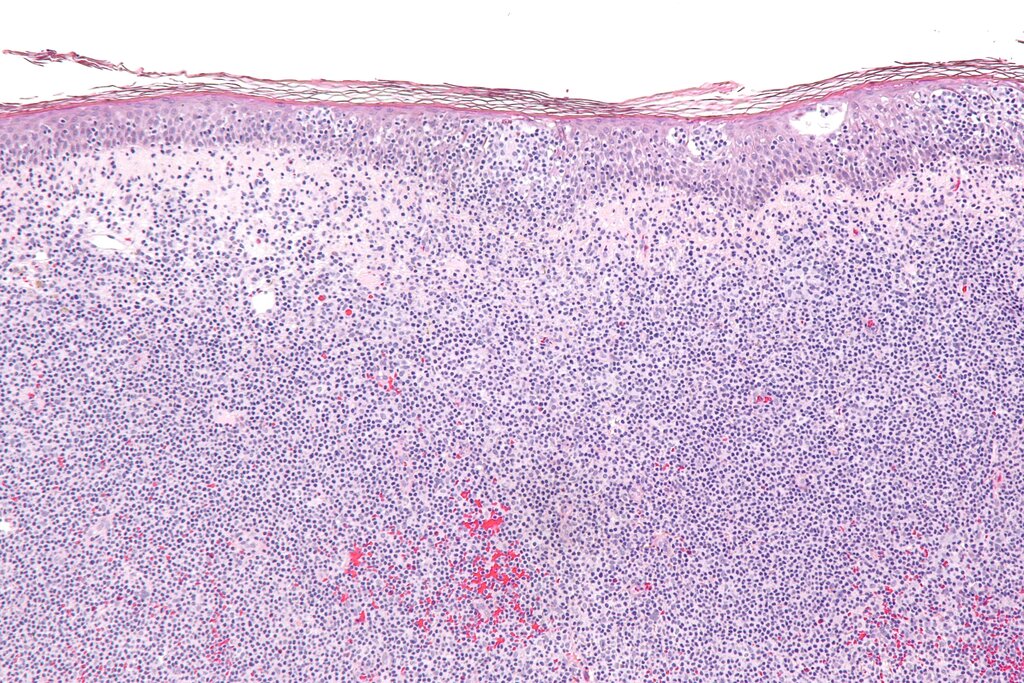
Type C presents the most alarming histologic picture. It is characterized by nodules or sheets of large, atypical CD30+ cells with minimal accompanying inflammatory infiltrate. This pattern can be indistinguishable from primary cutaneous anaplastic large cell lymphoma (pcALCL). However, the defining difference is clinical behavior. Despite the aggressive appearance under the microscope, Type C lesions follow the typical LyP course of spontaneous regression. This underscores the rule that LyP is a clinicopathologic diagnosis; histology alone is insufficient.
Emerging Variants: Types D, E, and F
The newer subtypes have refined our understanding of LyP's diversity. Type D is a striking variant marked by a dense, epidermotropic infiltrate of atypical small- to medium-sized lymphocytes. What sets it apart is its immunophenotype: the atypical cells are predominantly cytotoxic CD8+ T-cells. This presentation can simulate aggressive epidermotropic CD8+ cytotoxic T-cell lymphoma, a diagnosis with a much graver prognosis. Clinically, Type D may present with hemorrhagic or crusted papules.
Type E is defined by its angiocentric and angioinvasive nature. Histology reveals an infiltrate of atypical CD30+ lymphocytes centered on and often destroying blood vessel walls. This vascular damage leads to ischemic necrosis, which manifests clinically as ulcerative, eschar-like lesions that can leave scars. Like Type D, the atypical cells in Type E are often CD8+, and a subset may also express CD56, a marker sometimes associated with more aggressive lymphomas. The clinical presentation of deep necrotic ulcers is a key clue.
Finally, Type F introduces a specific genetic marker into the classification. This subtype is characterized by a follicular-based papular eruption. Histologically, it shows a perifollicular and intrafollicular infiltrate of atypical cells. The key feature is the immunophenotype: the neoplastic cells are CD4+ and show a (Tfh) profile, expressing markers like PD-1, BCL6, and CXCL13. Genetically, Type F is associated with a translocation involving the DUSP22-IRF4 locus on chromosome 6p25.3. This same translocation is found in a subset of pcALCL, highlighting the shared genetic pathways among CD30+ lymphoproliferative disorders.
| Subtype | Key Histologic Feature | Predominant Phenotype | Clinical Appearance |
|---|---|---|---|
| Type A | Wedge-shaped mixed infiltrate | CD4+, CD30+ | Papules, nodules |
| Type B | Epidermotropic, MF-like | CD4+, CD30+/- | Scaly papules, patches |
| Type C | Sheets of large atypical cells | CD4+, CD30+ | Nodules, tumors |
| Type D | Epidermotropic infiltrate | CD8+, CD30+ | Hemorrhagic papules |
| Type E | Angiocentric/Angioinvasive | CD8+, CD30+ | Necrotic ulcers, eschars |
| Type F | Perifollicular infiltrate | CD4+, Tfh profile, CD30+ | Follicular papules |
Distinguishing between these subtypes is more than an academic exercise. It sharpens our diagnostic accuracy, helps differentiate LyP from more aggressive lymphomas, and provides a more nuanced understanding of the patient's specific disease expression. The correlation between the microscopic findings and the clinical lesions on the patient’s skin remains the cornerstone of diagnosing all forms of LyP.
What is the defining clinical characteristic of Lymphomatoid Papulosis (LyP) that distinguishes it from more aggressive lymphomas like pcALCL, despite sometimes having histologically identical features?
A patient presents with scaly papules and patches. A biopsy reveals a band-like infiltrate in the papillary dermis with prominent epidermotropism of atypical lymphocytes with cerebriform nuclei. Only a few scattered cells are positive for CD30. Which LyP subtype is most likely?