Knee Replacement Surgery Explained
Knee Anatomy
The Knee's Bony Framework
The knee is where three major bones meet: the femur, the tibia, and the patella. It’s the largest joint in your body, and it has to handle a lot of stress from walking, running, and jumping.
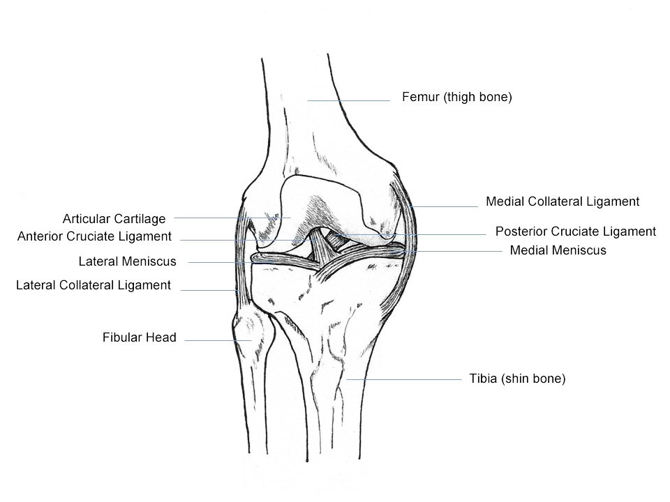
Your femur, or thigh bone, is the long bone that runs from your hip to your knee. Its bottom end is shaped into two rounded knobs called condyles, which rest on top of the tibia. The tibia, or shin bone, is the larger of the two bones in your lower leg. Its flat upper surface, the tibial plateau, forms the bottom half of the knee joint.
The third bone is the patella, commonly known as the kneecap.
Patella
noun
A small, flat, triangular bone that sits in front of the knee joint.
Connecting and Stabilizing
Bones alone can't form a stable joint. That’s where ligaments and cartilage come in. Ligaments are tough, elastic bands of tissue that connect bones to each other, acting like strong ropes to keep your knee from moving in the wrong directions.
The knee has four primary ligaments that provide most of its stability. They can be divided into two groups.
First are the collateral ligaments, found on the sides of your knee. The Medial Collateral Ligament (MCL) is on the inside, and the Lateral Collateral Ligament (LCL) is on the outside. They control the sideways motion of your knee and brace it against unusual movements.
The second group is the cruciate ligaments, which are found inside the knee joint. They cross each other to form an “X,” with the Anterior Cruciate Ligament (ACL) in front and the Posterior Cruciate Ligament (PCL) in back. The ACL stops the tibia from sliding out in front of the femur, while the PCL prevents the tibia from moving too far backward.
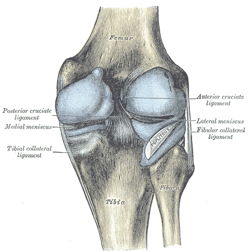
Between the femur and tibia are two C-shaped pieces of cartilage called the menisci. Each knee has a medial meniscus on the inside and a lateral meniscus on the outside. They act as shock absorbers, cushioning the joint and helping to distribute weight evenly. Without them, the bones would grind against each other.
Movement and Lubrication
Muscles and tendons work together to move the knee joint. Tendons are similar to ligaments, but they connect muscles to bones. The largest tendon in the knee is the patellar tendon, which covers the kneecap and connects the large quadriceps muscle in your thigh to the top of your tibia. When your quadriceps contract, they pull on the patellar tendon to straighten your leg.
“Not only does the knee have the two major leg bones, the femur and the tibia,” says Bhanu Ramaswamy, physio and visiting fellow at Sheffield Hallam University, “but you’ve got the kneecap, cartilage, tendons and ligaments, and any changes in those will make a knee prone to becoming more stiff or inflamed.”
To ensure all these moving parts work smoothly, the knee joint is enclosed in a capsule filled with synovial fluid. This thick, slippery liquid acts like oil in an engine, lubricating the joint to reduce friction and allow for smooth, painless motion. It also nourishes the cartilage, which doesn't have its own blood supply.
Ready to test your knowledge? Let's see what you've learned about the knee's structure.
Which of the following bones is NOT part of the knee joint?
What is the primary function of the menisci?
Understanding how these bones, ligaments, and fluids work together is the first step in learning about knee health and joint replacement.