Knee Pain Explained
Knee Anatomy
The Knee's Core Structure
The knee is where three major bones meet: the femur (thigh bone), the tibia (shin bone), and the patella (kneecap). The femur is the long bone in your upper leg, and its lower end flares out into two rounded knobs called condyles. These condyles rest on the flat upper surface of the tibia, known as the tibial plateau.
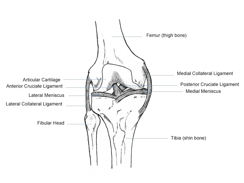
The patella, or kneecap, is a small, triangular bone that sits in front of the knee joint. It slides in a groove on the femur as you bend and straighten your leg. Its main job is to increase the leverage of the quadriceps muscles, making your leg extension more powerful.
Cushioning and Support
The ends of the femur and tibia, along with the back of the patella, are covered with a smooth, slippery substance called articular cartilage. This cartilage allows the bones to glide over each other with very little friction. It's like the ice on a skating rink, ensuring smooth movement.
Between the femur and tibia are two C-shaped pieces of tough, rubbery cartilage called menisci. You have a medial meniscus on the inner side of your knee and a lateral meniscus on the outer side. They act as shock absorbers, spreading out the force from your body weight and providing stability to the joint.
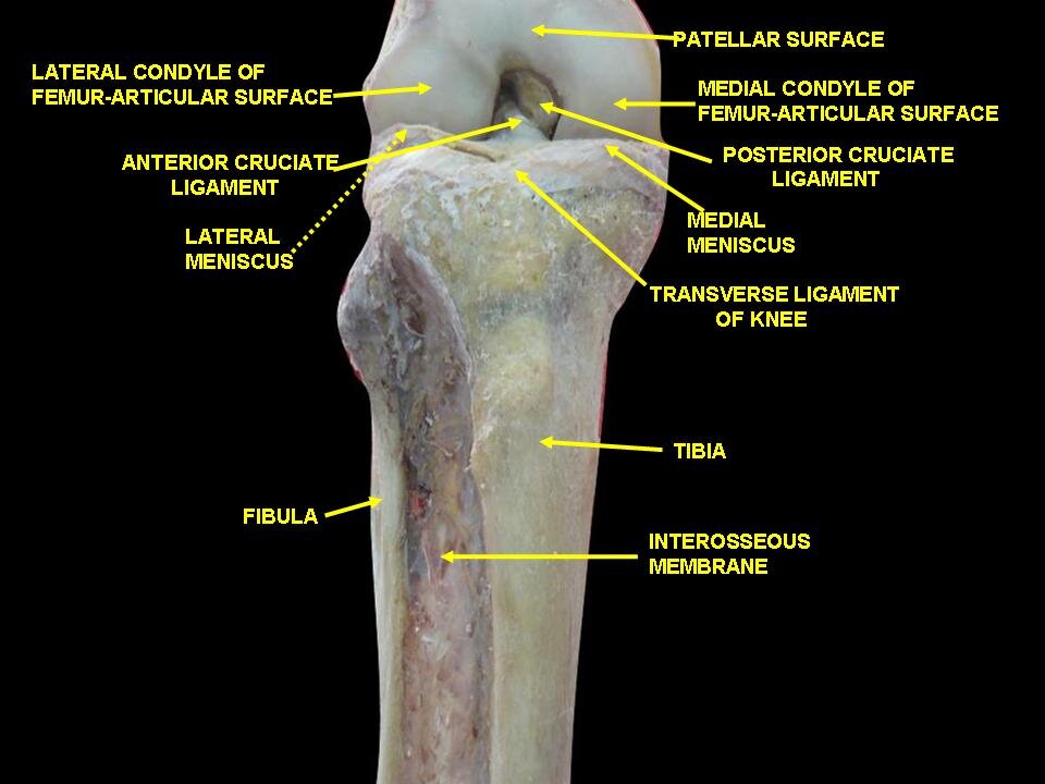
meniscus
noun
A crescent-shaped fibrocartilaginous structure that, in contrast to an articular disk, only partly divides a joint cavity.
The entire knee joint is enclosed in a fluid-filled sac called the joint capsule. The inner lining of this capsule, the synovial membrane, produces synovial fluid. This viscous fluid lubricates the joint, further reducing friction and nourishing the cartilage.
The Stabilizing Ligaments
Ligaments are strong, fibrous bands that connect bones to other bones. The knee has four main ligaments that provide its stability, acting like strong ropes to hold the joint together and control its motion.
The two collateral ligaments are on the sides of your knee. The Medial Collateral Ligament (MCL) runs along the inside of the knee and prevents it from bending inward. The Lateral Collateral Ligament (LCL) is on the outside and prevents the knee from bending outward.
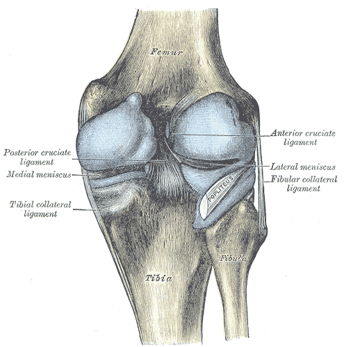
Inside the joint capsule are the cruciate ligaments, which cross each other in an "X" formation. The Anterior Cruciate Ligament (ACL) is at the front and prevents the tibia from sliding too far forward relative to the femur. The Posterior Cruciate Ligament (PCL) is at the back and stops the tibia from moving too far backward.
Together, the four main ligaments—MCL, LCL, ACL, and PCL—are the primary stabilizers of the knee, controlling its front-to-back and side-to-side movements.
Muscles, Tendons, and Movement
While ligaments provide stability, muscles and tendons provide movement. Tendons are tough cords that connect muscle to bone.
The quadriceps are a group of four large muscles on the front of your thigh. They come together to form the quadriceps tendon, which attaches to the top of the patella. This tendon continues over the patella and becomes the patellar tendon (sometimes called the patellar ligament), which then attaches to the tibia. When you contract your quadriceps, they pull on this tendon system to straighten your leg.
On the back of your thigh are the hamstrings, a group of three muscles responsible for bending your knee. Their tendons attach to the back of the tibia and the fibula (the smaller bone running alongside the tibia).
The knee's movement is a sophisticated combination of rolling and gliding. As you bend your knee from a straight position, the femoral condyles first roll and then glide on the tibial plateau. This complex motion, guided by the ligaments and powered by the muscles, allows for a large range of motion while maintaining stability.
Now, let's test your understanding of the knee's anatomy.
What is the primary function of the menisci in the knee joint?
The hamstring muscles are responsible for straightening the leg at the knee joint.
Understanding these components is the first step toward understanding how the knee functions and what can go wrong. Each part plays a critical role in allowing you to walk, run, and jump.