Introduction to Microscopy
Introduction to Microscopy
Seeing the Unseen
At its heart, a microscope is a tool that bends light to make small things look bigger. If you've ever used a magnifying glass to look at an insect or the fine print on a label, you've used a simple microscope. It uses a curved piece of glass, called a lens, to bend light rays and trick your eyes into seeing a larger version of the object.
Lenses are the key. By carefully shaping glass, we can control the path of light that passes through it. A single convex lens, thicker in the middle than at the edges, can magnify an object. Modern microscopes use a series of lenses working together to create a highly magnified and detailed image.
This focusing ability is the foundation of microscopy. By placing a specimen near the lens, we can capture the light coming from it and spread it out to form a much larger, virtual image for our eyes to see.
Magnification vs. Resolution
When we talk about a microscope's power, two terms are crucial: magnification and resolution. They sound similar, but they describe very different things.
Magnification
noun
The degree to which an object's apparent size is enlarged.
Magnification is simply how much bigger the microscope makes the object appear. A 100x magnification makes the object look 100 times larger than its actual size. But making something bigger isn't always enough. You also need clarity.
Resolution
noun
The ability to distinguish between two separate points. It is a measure of image clarity.
Resolution, or resolving power, is what allows us to see fine details. Imagine a low-quality photo on your computer. You can magnify it, making the pixels huge, but the image just becomes a blurry block. You haven't gained any new detail. High resolution means the microscope can distinguish between two points that are very close together, revealing them as separate entities instead of a single blur.
Magnification makes things look bigger. Resolution makes them look sharper.
Light vs. Electrons
The two fundamental types of microscopes are defined by what they use to "see" the specimen: light microscopes and electron microscopes.
A light microscope works just like the name suggests. It shines visible light through a specimen, and a system of lenses magnifies the image. This is the classic microscope you might picture from a science class. However, the resolution of light microscopes is limited by the properties of light itself. Because light travels in waves, we can't use it to clearly see things that are smaller than its wavelength.
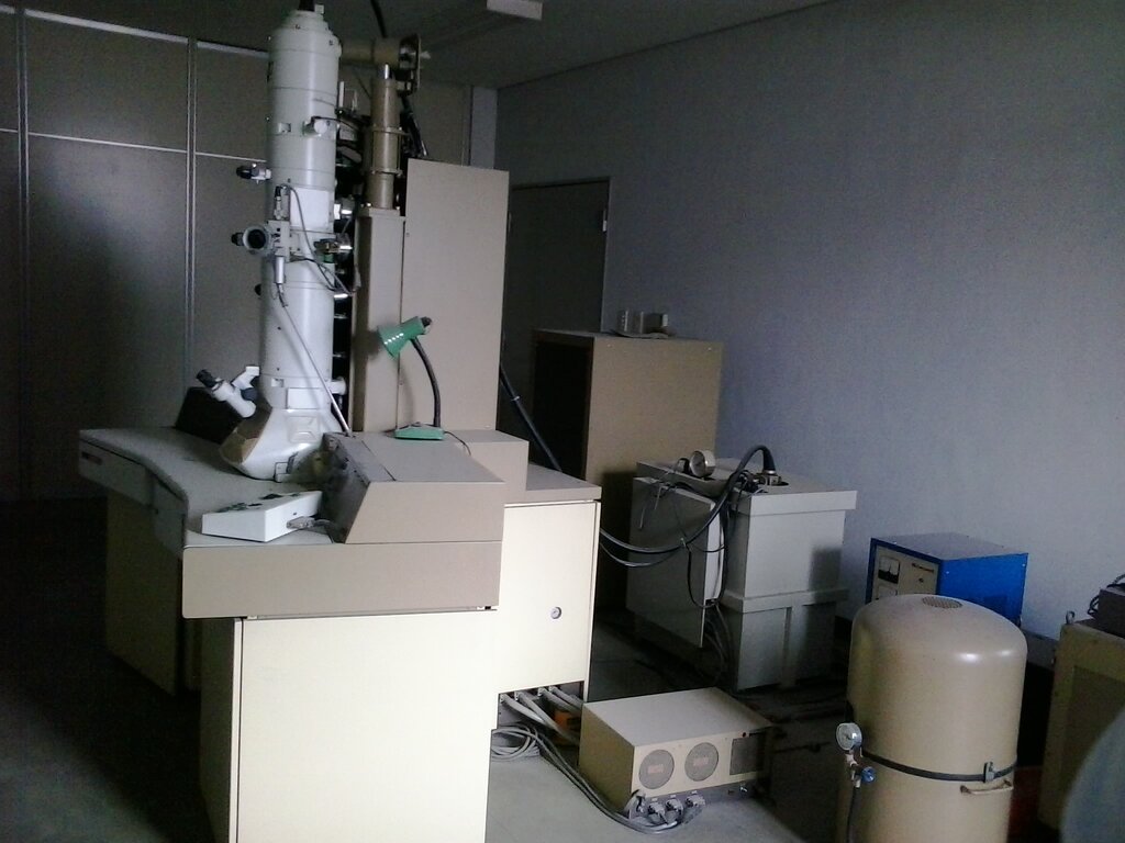
To see even smaller things, scientists needed something with a much shorter wavelength. The solution was the electron.
Electron microscopes use a beam of electrons instead of a beam of light. Electrons also behave like waves, but their wavelengths can be over 1,000 times shorter than visible light. This dramatically shorter wavelength is the secret to their power. It allows them to resolve incredibly small details, far beyond the reach of any light microscope.
Instead of glass lenses, electron microscopes use powerful electromagnets to focus the electron beam. By controlling these magnetic fields, they can create highly magnified and resolved images of everything from a virus particle to the surface of a metal atom.
What is the primary function of a lens in a microscope?
A scientist is viewing a sample at 400x magnification, but cannot distinguish two very small structures that are close together. The image is large, but blurry. What is the main issue?
Understanding these core principles of lenses, magnification, resolution, and the difference between light and electrons provides the foundation for exploring the incredible world of the very small.