Introduction to Cardiology
Heart Anatomy
The Four Chambers
Think of the heart as a muscular, four-room house. The top two rooms are called atria, and they act as receiving chambers for blood coming into the heart. The bottom two rooms are the ventricles, which are powerful pumps that send blood out of the heart.
The heart is divided into a right side and a left side by a muscular wall called the septum. This division is crucial because each side handles a different type of blood. The right side of the heart deals with deoxygenated blood (blood that has already delivered oxygen to the body's tissues), while the left side manages oxygenated blood (blood that has just picked up fresh oxygen from the lungs).
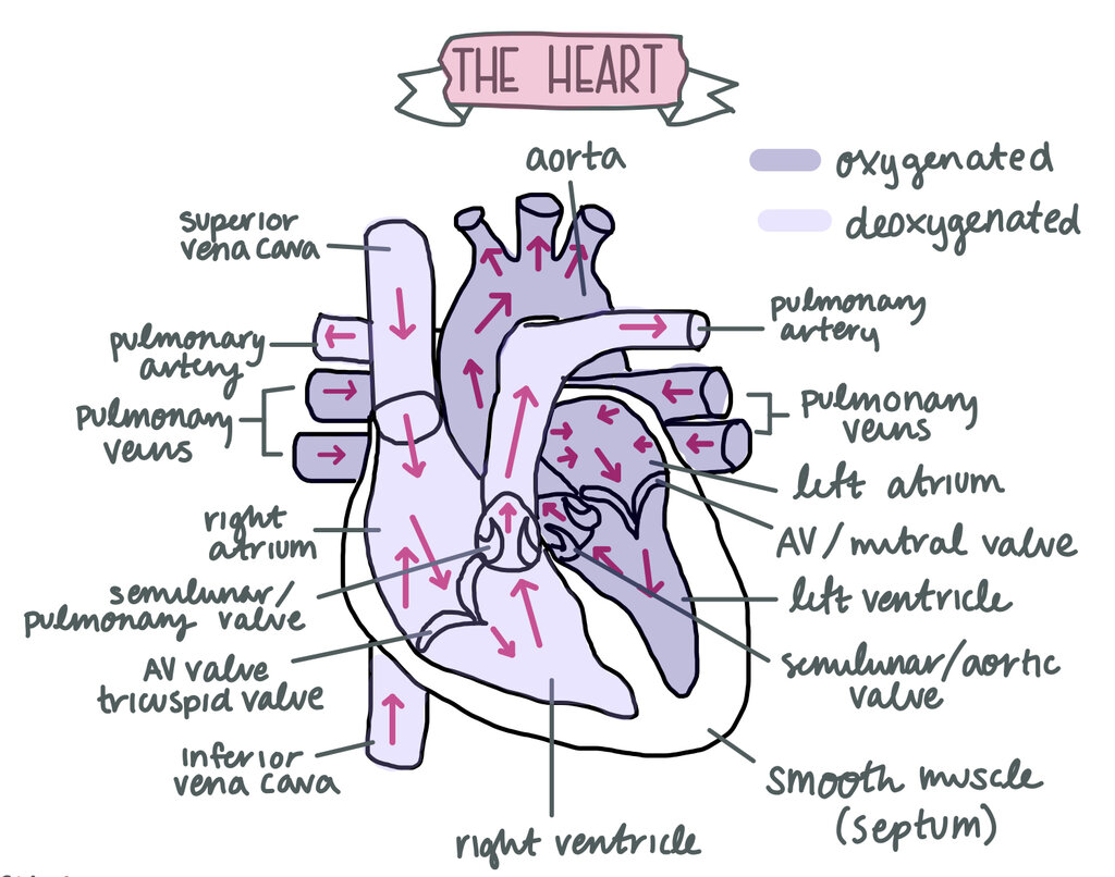
Let's walk through the rooms:
- Right Atrium: This chamber receives deoxygenated blood from the body through two large veins, the superior and inferior vena cava.
- Right Ventricle: When the right atrium is full, it pushes the blood into the right ventricle below it. This ventricle's job is to pump that deoxygenated blood to the lungs.
- Left Atrium: After the blood picks up oxygen in the lungs, it returns to the heart, entering the left atrium.
- Left Ventricle: From the left atrium, the now oxygen-rich blood moves into the left ventricle. This is the heart's strongest chamber. Its thick, muscular walls give it the power to pump oxygenated blood out to the entire body.
The Heart's Doors
To keep blood flowing in the correct direction, the heart has four valves. These act like one-way doors, opening to let blood pass through and then snapping shut to prevent it from flowing backward. There are two main types: atrioventricular (AV) valves and semilunar valves.
The AV valves are located between the atria and the ventricles, while the semilunar valves are found at the exits of the ventricles.
The tricuspid valve is the AV valve on the right side of the heart, between the right atrium and right ventricle. It has three small flaps, or cusps, which is where it gets its name.
The mitral valve (also called the bicuspid valve) is the AV valve on the left side, separating the left atrium and left ventricle. It has two cusps.
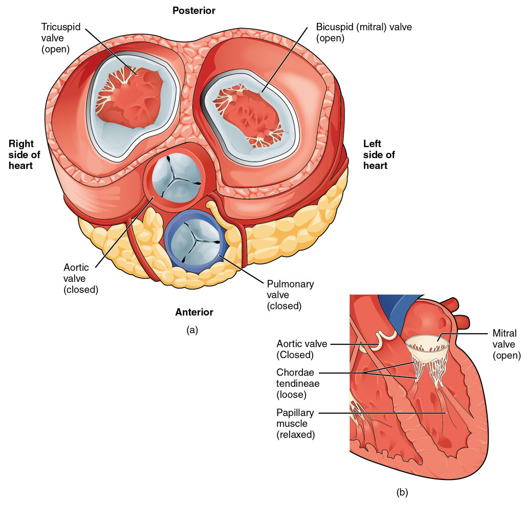
The two semilunar valves guard the arteries leaving the heart.
- The pulmonary valve is positioned between the right ventricle and the pulmonary artery. It opens to allow deoxygenated blood to be pumped to the lungs.
- The aortic valve sits between the left ventricle and the aorta. It opens to let oxygen-rich blood flow out to the rest of the body.
Major Highways
Several large blood vessels are connected directly to the heart, serving as the main highways for blood entering and leaving. These are the great vessels.
Bringing blood to the heart:
- Superior and Inferior Vena Cava: These are the two largest veins in the body. The superior vena cava collects deoxygenated blood from the upper half of the body (head, neck, arms), while the inferior vena cava collects it from the lower half. Both empty into the right atrium.
- Pulmonary Veins: Unlike most veins, these carry oxygen-rich blood. They transport blood from the lungs back to the left atrium.
Sending blood away from the heart:
- Pulmonary Artery: This large artery carries deoxygenated blood from the right ventricle to the lungs to pick up oxygen. It's a rare example of an artery that carries deoxygenated blood.
- Aorta: The largest artery in the body. It carries oxygenated blood from the powerful left ventricle and distributes it to every part of the body, from your brain to your toes.
Now let's test your knowledge of the heart's structure.
What is the primary function of the heart's ventricles?
Which chamber of the heart directly receives deoxygenated blood from the body through the superior and inferior vena cava?
Understanding these basic components—the chambers, valves, and major vessels—is the foundation for learning how the entire cardiovascular system functions.