Interpreting Echocardiograms
Introduction to Echocardiography
Seeing the Heart with Sound
An echocardiogram is essentially an ultrasound of the heart. It’s a safe, non-invasive way for doctors to get a detailed look at this vital organ without any radiation or surgery. By using sound waves, this technique creates dynamic, real-time images of the heart's chambers, valves, walls, and blood vessels.
Echocardiography is the most widely used cardiac imaging modality, capturing ultrasound video data to assess cardiac structure and function.
This allows clinicians to see how the heart is beating and pumping blood, helping them diagnose a wide range of conditions, from valve problems to damage from a heart attack.
The Physics of Ultrasound
The science behind echocardiography is based on the simple principle of sound waves and echoes. The machine uses a type of sound called ultrasound, which has a frequency higher than the upper limit of human hearing. Think of it like a bat using echolocation to navigate in the dark.
A specialized probe sends short pulses of these high-frequency sound waves into the body. As the waves travel, they encounter different types of tissue, like heart muscle, blood, and bone. Each tissue reflects the sound waves differently.
Dense structures, like bone or heart valves, reflect a lot of sound and appear bright white on the screen. Less dense materials, like blood, let most of the waves pass through and appear dark.
The same probe that sends the signals also listens for the returning echoes. The machine measures the time it takes for these echoes to return and their strength. It then uses this information to build a live, moving picture of the heart. Some sound waves will get weaker or absorbed as they travel deeper, a process known as attenuation, which the machine accounts for when creating the image.
The Tools of the Trade
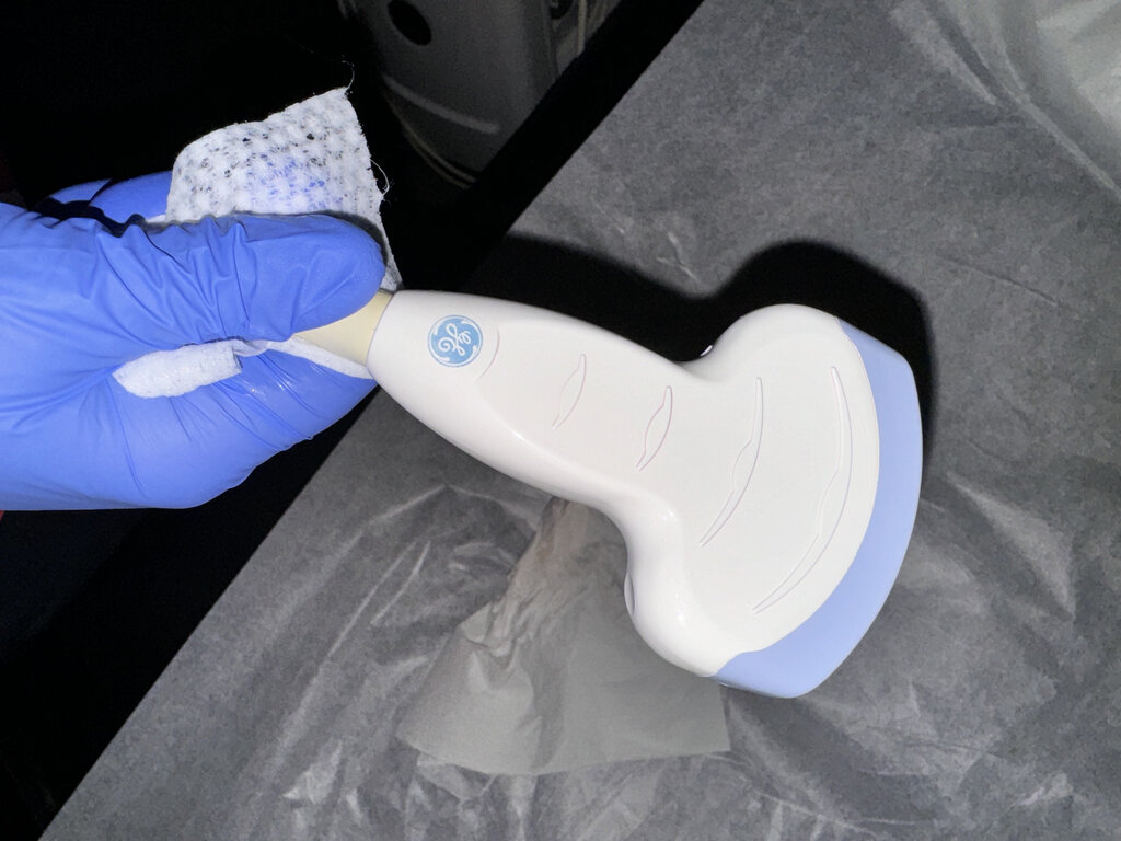
The main piece of equipment for an echocardiogram is the ultrasound machine itself. It consists of a computer console with a display screen and a handheld device called a transducer.
transducer
noun
A device that converts energy from one form to another. In ultrasound, it converts electrical energy into sound waves and the returning sound waves (echoes) back into electrical signals.
The transducer is the key component that touches the patient's body. Inside it are piezoelectric crystals that vibrate when an electric current is applied, creating the ultrasound waves. When the echoes return, they hit these same crystals, causing them to vibrate and generate an electric current. This signal is sent back to the main console, which processes it into the images seen on the screen.
Different Imaging Modes
Echocardiography isn't just one type of picture. Sonographers use several different modes to gather specific information about the heart.
| Mode | Description | Primary Use |
|---|---|---|
| 2D Mode | Creates a live, two-dimensional, cross-sectional "slice" of the heart. | Visualizing heart structures, chambers, and valves in real-time. |
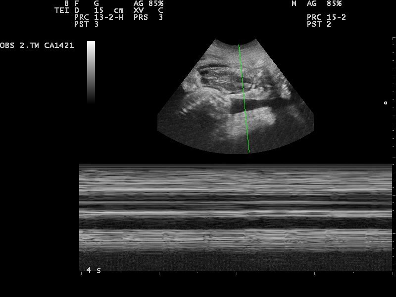
| M-Mode | Takes a single line from the 2D image and displays its motion over time on a graph. | Measuring chamber dimensions and timing cardiac events precisely. |
| Doppler | Measures the speed and direction of blood flow within the heart. | Detecting leaks in valves, checking for blockages, and assessing blood pressure inside the heart. |
Doppler ultrasound works on the same principle as a passing ambulance siren: the pitch of the sound changes depending on whether it's moving toward you or away from you. The machine uses this change in the sound waves' frequency to create a map of blood flow, often displayed with colors to show direction (Color Doppler) or as a graph to show velocity (Pulsed-Wave or Continuous-Wave Doppler).
Pros and Cons
Like any medical procedure, echocardiography has its strengths and weaknesses.
Advantages
- Safe: It uses no ionizing radiation.
- Real-Time: It provides immediate, live images of the heart in motion.
- Accessible: The equipment is widely available and less expensive than other imaging methods like MRI or CT scans.
- Portable: Machines can be brought to a patient's bedside, making it useful in intensive care and emergency situations.
Limitations
- Operator Dependent: The quality of the exam heavily relies on the skill of the person performing it.
- Patient Factors: A clear view can be difficult to obtain in patients with obesity, severe lung disease, or certain chest wall shapes.
- Limited View: Ribs and lungs can sometimes block the sound waves, creating acoustic shadows that hide parts of the heart.
Despite these limitations, echocardiography remains a cornerstone of cardiac diagnosis due to its safety, versatility, and power to visualize the living, beating heart.
What is the fundamental principle used by an echocardiogram to create images of the heart?
Which component inside the transducer is responsible for both generating the ultrasound waves and detecting their returning echoes?