Immunohistochemistry and Histopathology Diagnostics
Antigen Retrieval Mastery
The Formalin Fix
Formalin fixation is the gold standard for preserving tissue structure. It works by creating a web of chemical bonds, called , that lock proteins and other molecules in place. This process essentially freezes the cellular architecture, providing a stable snapshot for analysis. However, this excellent preservation comes at a cost. The very cross-links that stabilize the tissue also mask the antigenic sites, or epitopes, that antibodies need to recognize. It's like covering a keyhole with a thick layer of plaster. The keyhole is still there, but the key, our primary antibody, can no longer fit. To make the epitope accessible again, we must break these cross-links without destroying the tissue itself. This crucial step is called antigen retrieval.
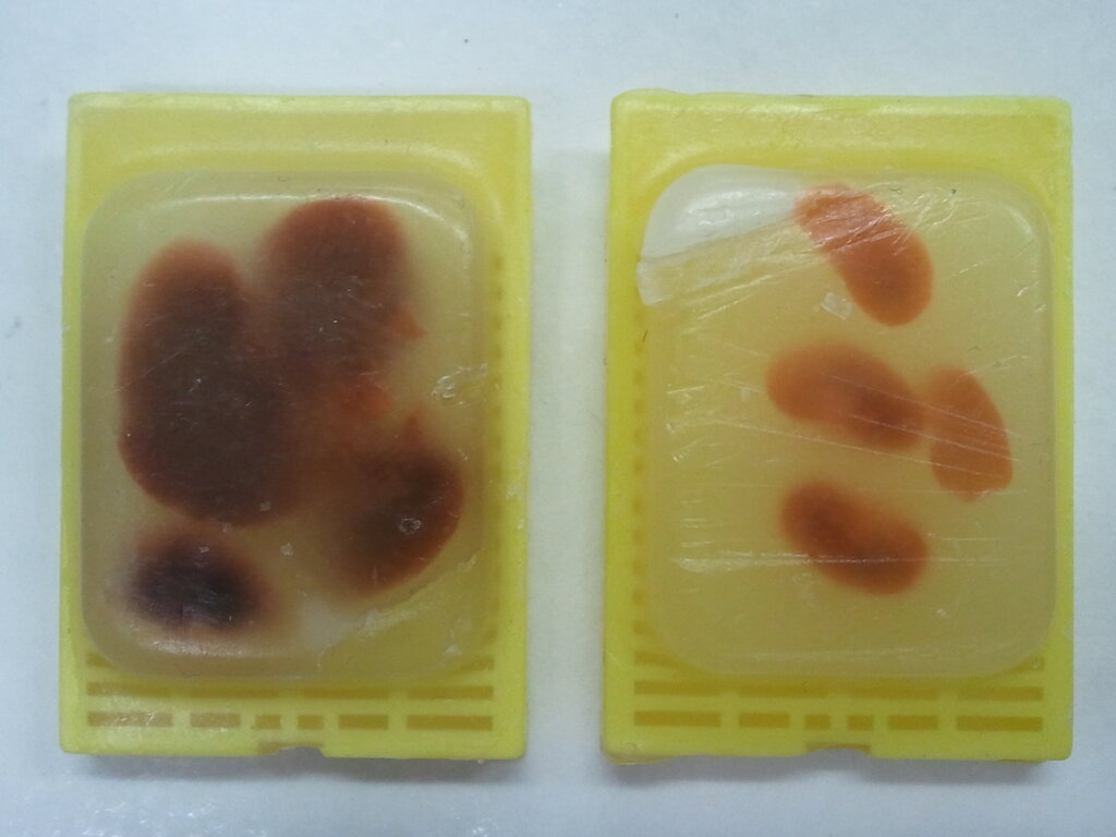
The goal is to reverse the masking effect of fixation just enough for the antibody to bind, while keeping the overall tissue structure intact. This delicate balance is the core of successful immunohistochemistry. There are two primary philosophies for achieving this: breaking the bonds with heat or gently chewing them away with enzymes.
Heat vs. Enzymes
The two main strategies for antigen retrieval are Heat-Induced Epitope Retrieval (HIER) and Proteolysis-Induced Epitope Retrieval (PIER). They approach the problem from different angles.
HIER is the most common method used today. It involves heating the tissue sections in a specific buffer solution. The heat provides the energy needed to hydrolyze and break the methylene bridges. Think of it as carefully melting the plaster from our keyhole analogy. It's a physical process driven by temperature and time.
PIER, on the other hand, uses enzymes like proteinase K, trypsin, or pepsin to carefully digest some of the proteins that are blocking the epitope. This is a more targeted biochemical approach, like using a tiny chisel to chip away the plaster. The key is to let the enzyme work just long enough to expose the target without degrading the entire tissue structure. PIER is often preferred for epitopes located on the cell surface or within the cytoplasm that are particularly sensitive to heat.
| Feature | Heat-Induced Epitope Retrieval (HIER) | Proteolysis-Induced Epitope Retrieval (PIER) |
|---|---|---|
| Mechanism | Breaks cross-links using heat energy | Uses enzymes to digest masking proteins |
| Common For | Nuclear and cytoplasmic antigens | Cytoplasmic and membrane-bound antigens |
| Control | Temperature, time, buffer pH | Enzyme concentration, time, temperature |
| Risk | Tissue detachment, morphology damage from heat | Over-digestion, loss of target antigen |
| Reproducibility | Generally higher and more consistent | Can be variable if conditions aren't precise |
Choosing between HIER and PIER depends on the specific antibody and the location of your target antigen. Most modern antibody datasheets recommend a specific retrieval method that has been optimized by the manufacturer. HIER has become the default for many labs due to its high success rate and reproducibility, especially with automated systems.
Optimizing HIER
Successful HIER depends on three interacting variables: buffer pH, temperature, and time. The pH of the retrieval buffer is arguably the most critical factor. Two buffers cover the vast majority of HIER protocols:
- Citrate Buffer (pH 6.0): This acidic buffer is a versatile workhorse and a good starting point for many antigens. It's effective for a wide range of cytoplasmic and some nuclear proteins.
- Tris-EDTA Buffer (pH 9.0): This alkaline buffer provides a more robust retrieval, especially for difficult nuclear antigens or when a citrate buffer yields weak staining. The higher pH is more effective at breaking certain cross-links and restoring the antigen's native conformation.
The choice of buffer isn't arbitrary. It's based on the biochemical properties of the target epitope. The pH affects the charge of the amino acid residues on the protein, and the correct pH helps the epitope refold into a recognizable shape after the cross-links are broken.
As a rule of thumb: start with Citrate pH 6.0. If staining is weak or absent, especially for a nuclear target, switch to Tris-EDTA pH 9.0.
Temperature and time are a balancing act. The goal is to heat the slide just long and hot enough to unmask the epitope without boiling the tissue off the slide. A typical target is 95–100°C for 20–40 minutes. However, the heating method itself matters.
- Pressure Cookers & Steamers: Provide consistent, even heating and are very effective. They are a low-cost, high-throughput option.
- Microwaves: Fast and readily available, but can suffer from uneven heating and
, leading to inconsistent results if not carefully managed.
- Water Baths: Offer gentle, stable heating but are typically slower and operate at slightly lower maximum temperatures.
- Automated Stainers: The most reproducible option. These instruments have integrated retrieval modules that precisely control temperature, time, and buffer changes, eliminating many sources of human error.
Troubleshooting Retrieval
Even with an optimized protocol, problems can arise. Two common issues are poor morphology and tissue detachment.
Poor Morphology or Over-staining: If your tissue looks 'mushy' or 'eaten away,' especially after PIER, you've likely over-digested it. Reduce the enzyme concentration or incubation time. For HIER, excessive heat or time can also damage tissue. This often manifests as intense, non-specific background staining where the antibody sticks to damaged proteins.
Tissue Detachment: Seeing your precious section float off the slide is frustrating. This is often a sign of excessive heat. Ensure your slides are properly charged (adhesive) and consider reducing the temperature or time slightly. Aggressive boiling is a key culprit; a steady, high-temperature 'simmer' is what you're after. Some delicate tissues, like brain or skin, are more prone to detaching and may require gentler conditions.
Good laboratory practice requires establishing optimal antibody concentration and antigen retrieval and detection methods.
Mastering antigen retrieval is a foundational skill. It transforms a fixed piece of tissue from an inaccessible archive into a rich source of molecular information, paving the way for accurate and insightful analysis.
What is the primary purpose of antigen retrieval in immunohistochemistry?
The chemical bonds created by formalin fixation that mask antigenic sites are called __________.