ICU Monitoring and Ventilator Modes MCQs for Physiotherapists
Non-Invasive Monitoring
Monitoring Without Breaking the Skin
In the Intensive Care Unit (ICU), a patient's condition can change in an instant. Continuous monitoring is essential, but we don't always need to use invasive methods that puncture the skin or enter the body. Non-invasive monitoring gives us a constant stream of vital information safely and with less risk to the patient. These tools are the cornerstones of modern critical care, providing a window into the body's function second by second.
Vital signs are critical indicators of a patient's health status and are essential components of nursing assessments.
For physiotherapists, understanding these monitors is crucial. Your treatment decisions, from mobilization to respiratory interventions, depend on interpreting this data correctly. A dropping oxygen saturation level or an irregular heart rhythm can tell you to stop or modify your approach. Let's look at the three main types you'll encounter.
Pulse Oximetry
Pulse oximetry is one of the most common non-invasive techniques. You've likely seen the small clip placed on a patient's finger, toe, or earlobe. Its job is to estimate the amount of oxygen in the blood, a value called oxygen saturation, or .
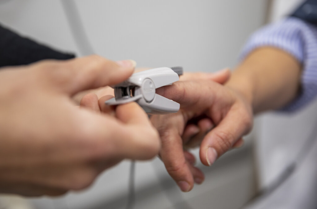
The device works by shining two types of light, red and infrared, through the tissue. Hemoglobin, the protein in red blood cells that carries oxygen, absorbs these two lights differently depending on whether it's carrying an oxygen molecule. Oxygenated hemoglobin absorbs more infrared light, while deoxygenated hemoglobin absorbs more red light. The sensor on the other side of the clip measures how much of each light passes through. By analyzing the ratio of light absorbed, the machine calculates the percentage of hemoglobin that is saturated with oxygen.
A healthy is typically 95-100%. A value below 90% is a cause for concern and indicates hypoxemia. However, readings can be inaccurate. Poor circulation to the limb, dark nail polish, excessive patient movement, or very bright ambient light can all interfere with the signal. Always correlate the reading with the patient's clinical presentation. Does the patient look short of breath? Are their lips or nail beds blue? If the reading doesn't match the patient, start troubleshooting.
A common issue is a poor waveform on the monitor. A strong, consistent waveform that matches the patient's heart rate suggests a reliable reading. If it's erratic or flat, reposition the probe or try a different location.
The Heart's Electrical Story
Electrocardiography, or ECG, tracks the electrical activity of the heart. In the ICU, this is typically done continuously using a 3-lead or 5-lead system. Small, sticky electrodes are placed on the patient's chest, and wires connect them to a monitor. This setup doesn't give the diagnostic detail of a full 12-lead ECG, but it's excellent for monitoring the heart's rate and rhythm in real-time.
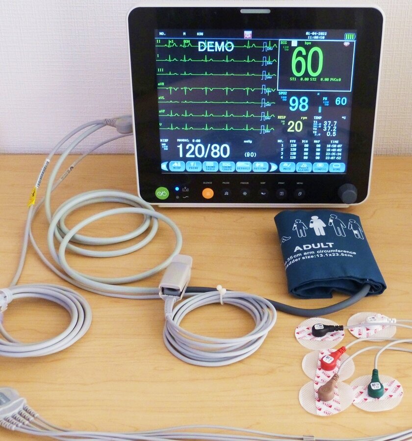
The monitor displays this electrical activity as a waveform. You'll primarily be watching for two things: the heart rate (how fast the heart is beating) and the rhythm (whether the beat is regular or irregular). For a physiotherapist, this is critical. An abnormally high heart rate (tachycardia) or low heart rate (bradycardia) during therapy might mean the patient isn't tolerating the activity. The appearance of new arrhythmias, like atrial fibrillation, is a red flag that requires immediate attention from the medical team.
| Common ICU Arrhythmias | Key Feature |
|---|---|
| Sinus Tachycardia | Normal rhythm, but rate > 100 bpm. Often due to pain, fever, or exertion. |
| Sinus Bradycardia | Normal rhythm, but rate < 60 bpm. Can be normal in athletes, but concerning in sick patients. |
| Atrial Fibrillation (AFib) | Irregularly irregular rhythm with no clear P waves. Increases stroke risk. |
| Ventricular Tachycardia (VTach) | A fast, wide-complex rhythm originating in the ventricles. A medical emergency. |
Troubleshooting ECG monitoring often involves the electrodes. If the signal is poor or filled with static (called 'artifact'), an electrode may have dried out or fallen off. Patient movement, especially shivering, can also create significant artifact. Check that all connections are secure and that the patient's skin is clean and dry where the electrodes are placed.
Blood Pressure Without the Puncture
Non-invasive blood pressure (NIBP) monitoring is usually done with an automated cuff, typically placed on the upper arm. The monitor is set to inflate the cuff at regular intervals, such as every 15 minutes or every hour, to measure the patient's blood pressure.
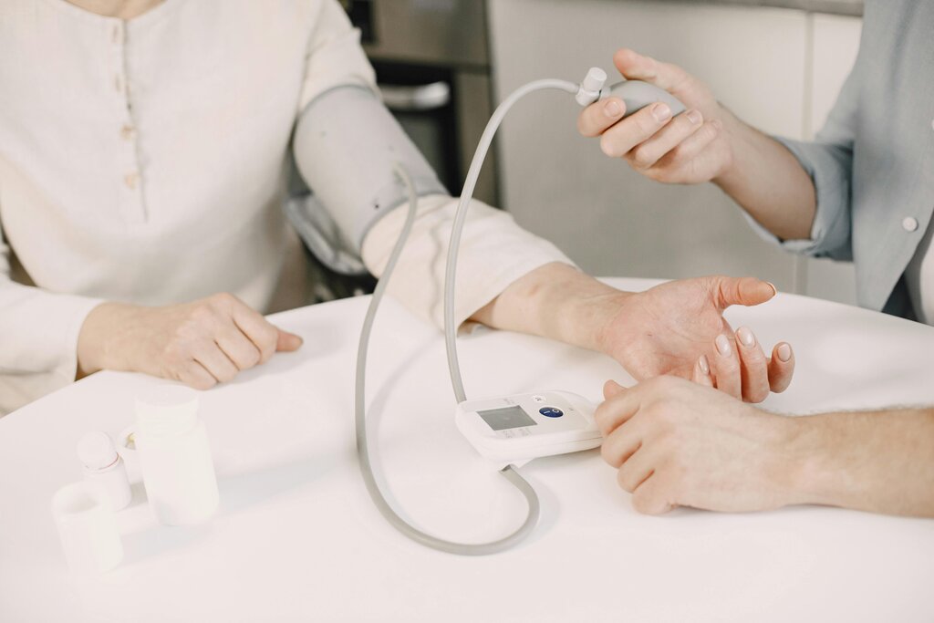
The machine uses an oscillometric method. As the cuff inflates and then slowly deflates, a sensor detects oscillations in the artery wall caused by blood flow. The point of maximum oscillation corresponds to the Mean Arterial Pressure (MAP). The machine then uses an algorithm to calculate the systolic and diastolic pressures from this data.
A common problem with NIBP is selecting the wrong cuff size. A cuff that's too small will give a falsely high reading, while a cuff that's too large will give a falsely low one. The cuff's bladder should encircle about 80% of the arm's circumference. Also, be mindful of where you place the cuff. Avoid arms with IV lines, fistulas, or injuries. Frequent inflation can be uncomfortable for the patient and can even cause bruising, so don't set the interval to be more frequent than clinically necessary.
When you mobilize a patient, their blood pressure will naturally change. However, a sharp drop, especially if accompanied by dizziness or fainting, is a sign of orthostatic hypotension and indicates the patient isn't ready for that level of activity. Always check the blood pressure before, during, and after your interventions.
Ready to test your knowledge? This quiz will cover the key points of non-invasive monitoring.
A pulse oximeter estimates blood oxygen saturation () by measuring the differential absorption of what two types of light?
While mobilizing a patient in the ICU, you notice the ECG monitor is showing a lot of static, known as 'artifact', making the heart rhythm difficult to read. What is the most likely cause?
By mastering the interpretation of these non-invasive tools, you become a more effective and safer clinician in the complex ICU environment.