Hypertrophy Training Principles
Muscle Anatomy
The Blueprint of a Muscle
Skeletal muscles, the engines that power our movements, are intricate structures. They aren't just single chunks of tissue. Instead, they are organized in a clever bundle-within-a-bundle system, starting from the entire muscle and narrowing down to microscopic fibers.
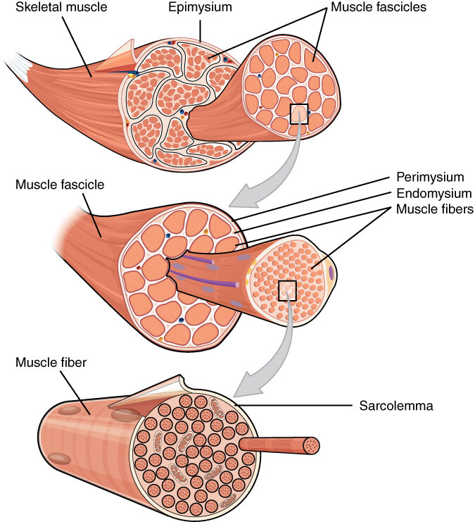
At the core of this structure is the muscle fiber, which is actually a single, long muscle cell. These fibers are the fundamental units responsible for contraction. Each muscle you have, from the large ones in your legs to the small ones in your face, is made up of thousands of these fibers bundled together.
These bundles of muscle fibers are called fascicles. Think of it like a thick electrical cable, where the entire cable is the muscle, and inside are many smaller, insulated wires. Each of those smaller wires is a fascicle, and inside each of those are the tiny copper filaments, the muscle fibers.
Two Types of Fibers
Not all muscle fibers are created equal. They are generally classified into two main types based on how quickly they contract and how they produce energy.
Type I fibers are known as slow-twitch fibers. They are built for endurance. They contract slowly but can work for long periods without fatiguing. These fibers are rich in mitochondria and myoglobin, which help them use oxygen to generate fuel efficiently. This makes them crucial for activities like marathon running or cycling.
Type II fibers are fast-twitch fibers, designed for powerful, explosive movements. They contract quickly but also tire quickly. These fibers are larger and generate more force than Type I fibers, making them essential for sprinting, jumping, and heavy weightlifting. They rely more on anaerobic metabolism, which doesn't require oxygen, for their quick bursts of energy.
| Feature | Type I Fibers (Slow-Twitch) | Type II Fibers (Fast-Twitch) |
|---|---|---|
| Contraction Speed | Slow | Fast |
| Fatigue Rate | Slow | Fast |
| Primary Activity | Endurance (e.g., running) | Power (e.g., sprinting) |
| Force Production | Low | High |
| Energy Source | Oxygen (Aerobic) | Glycogen (Anaerobic) |
Most muscles in your body contain a mix of both Type I and Type II fibers. However, the exact ratio is determined by genetics and the primary function of the muscle. For example, postural muscles in your back, which work all day to keep you upright, have a higher concentration of endurance-focused Type I fibers.
The Connective Tissue Framework
This entire system of fibers and fascicles is held together and supported by layers of connective tissue. This tissue provides structure, separates different parts of the muscle, and creates pathways for nerves and blood vessels.
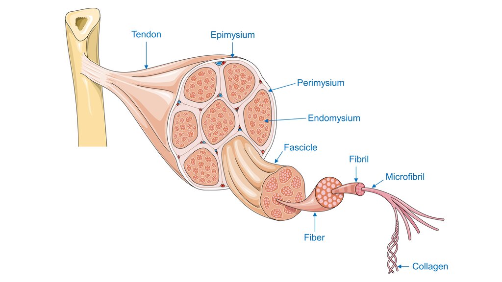
There are three main layers of this supportive tissue:
- Endomysium: The innermost layer, a delicate sheath that surrounds each individual muscle fiber.
- Perimysium: A thicker layer that wraps around a bundle of fibers to form a fascicle.
- Epimysium: The tough, outer layer that encases the entire muscle, separating it from surrounding tissues.
These three layers merge together at the ends of the muscle to form a strong, cord-like structure called a tendon. The tendon is what attaches the muscle to the bone, allowing the force of muscle contraction to create movement.
Now, let's test your knowledge of muscle structure and fiber types.
What is the correct hierarchical organization of a skeletal muscle, from the largest component to the smallest?
An elite marathon runner's leg muscles would likely have a higher concentration of which type of muscle fiber?
Understanding this basic anatomy is the first step. The way these fibers are organized and supported by connective tissue sets the stage for how they contract, perform, and ultimately, grow.