Human Locomotion and Muscular Physiology
Muscular Ultra-structure
Inside the Muscle Fiber
To understand how a muscle generates force, we need to zoom in. Way in. If you were to look inside a single muscle fiber, you'd find it's packed with thousands of smaller, cylindrical structures called myofibrils. These are the engines of the cell, running its entire length.
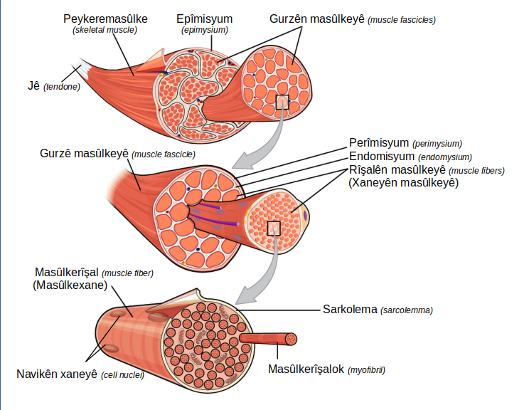
Each myofibril is a chain of repeating contractile units laid end-to-end. This fundamental unit is called the sarcomere. When a muscle contracts, it's because millions of these sarcomeres are shortening in unison. Their highly organized structure is what gives skeletal muscle its characteristic striped, or striated, appearance.
Anatomy of a Sarcomere
A single sarcomere is defined as the segment between two Z-lines, which are dense protein discs that act as anchors. Extending from these Z-lines are the thin filaments. In the center of the sarcomere are the thick filaments, held in place by another protein structure called the M-line.
This arrangement creates a distinct pattern of light and dark bands.
- A-bands: The dark bands, which represent the entire length of the thick filaments. The edges are darker because the thick and thin filaments overlap here.
- I-bands: The light bands, containing only thin filaments. These bands are bisected by the Z-line.
- H-zone: A paler region in the middle of the A-band where there are only thick filaments, with no thin filament overlap.
During contraction, the I-bands and H-zone shrink, while the A-band remains the same length. This is a key piece of evidence for how muscles work at a molecular level.
The Protein Players
The bands and lines of the sarcomere are formed by two main types of protein filaments: thick filaments and thin filaments. Their ability to interact is the basis of all muscle contraction.
Thick Filaments are primarily composed of a protein called myosin. Each myosin molecule has a long tail and two globular heads. Hundreds of these molecules are bundled together, with their tails forming the core of the filament and their heads projecting outwards.
These are the real workhorses. They can bind to the thin filaments and pivot, acting like tiny oars that pull the thin filaments toward the center of the sarcomere.
Thin Filaments are more complex. Their main component is the protein actin, which consists of individual globular subunits (G-actin) that link together to form a helical chain (F-actin). Each actin subunit has a binding site for a myosin head.
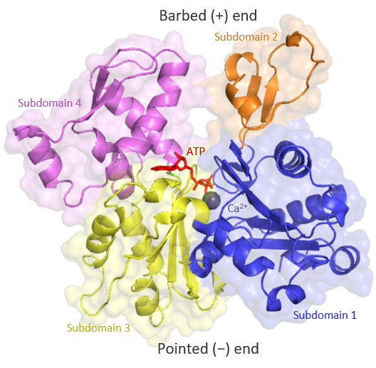
However, in a resting muscle, these binding sites are covered up by two regulatory proteins: tropomyosin and troponin.
- Tropomyosin: A long, fibrous protein that spirals around the actin chain, physically blocking the myosin-binding sites.
- Troponin: A smaller, complex protein attached to tropomyosin. It has a binding site for calcium ions ().
When calcium ions are released into the muscle fiber, they bind to troponin. This binding causes troponin to change shape, which in turn pulls the tropomyosin strand away from the actin binding sites. With the sites exposed, the myosin heads can now attach, and contraction can begin. These proteins act as the on/off switch for muscle activity.
Let's check your understanding of these microscopic structures.
What is the fundamental contractile unit of a myofibril, defined as the segment between two Z-lines?
During muscle contraction, which of the following remains the same length?
This intricate molecular architecture is the foundation of movement. Now that we've seen the parts, we can explore how they work together to make the muscle contract.