Human Anatomy and Clinical Physiology
Cardiorespiratory Integration
The Oxygen Transport Cascade
Your body's survival depends on a constant supply of oxygen. This delivery system, known as the oxygen transport cascade, describes the journey of oxygen from the air you breathe all the way to the mitochondria inside your cells, where it fuels energy production. It’s a multi-step process, and a drop in efficiency at any stage can have significant consequences.
The cascade begins with the partial pressure of oxygen in the atmosphere and ends with its consumption in the cell. Each step involves a pressure drop, which is what drives the oxygen forward. Think of it like a series of waterfalls; water only flows downhill, and oxygen only moves from an area of higher pressure to one of lower pressure. The key stages are:
- Inspired Air: Oxygen enters the lungs.
- Alveolar Gas: Oxygen crosses from the tiny air sacs (alveoli) into the blood.
- Arterial Blood: Oxygen-rich blood is pumped by the heart to the body.
- Capillaries: Oxygen leaves the blood and enters the tissues.
- Mitochondria: Oxygen is finally used by the cells to produce ATP, the body's energy currency.
Matching Airflow to Blood Flow
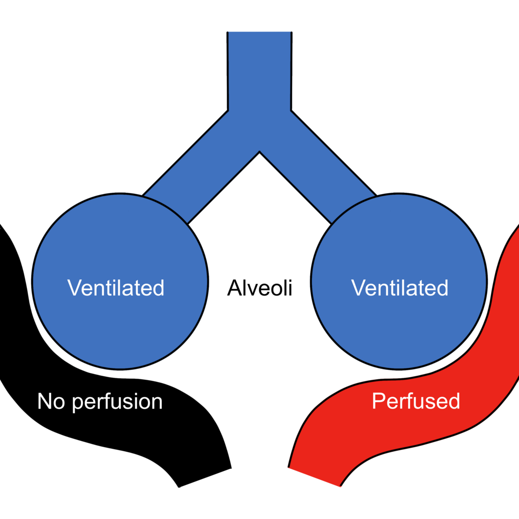
For gas exchange to be efficient, the air entering the alveoli must be matched with the blood flowing past them. This relationship is called the . An ideal V/Q ratio is around 0.8, meaning ventilation is slightly less than perfusion, but in a healthy lung, this ratio varies from top to bottom due to gravity. At the top (apex) of the lung, there is more air than blood (high V/Q), and at the bottom (base), there is more blood than air (low V/Q).
Your body actively tries to match V and Q. If an area of the lung is poorly ventilated, the blood vessels in that region constrict. This is called hypoxic pulmonary vasoconstriction. It's a smart reflex that diverts blood away from poorly oxygenated areas and toward areas that are working well. This shunts blood to where it can pick up oxygen, optimising the overall efficiency of the lung.
When the Systems Clash
The heart and lungs are so tightly linked that a problem in one often causes a problem in the other. This is known as cardiorespiratory interaction. A classic example is , which is right-sided heart failure brought on by chronic lung disease, such as Chronic Obstructive Pulmonary Disease (COPD).
In COPD, damaged lung tissue leads to chronic hypoxia. As we've learned, the body's response to low oxygen in the lungs is to constrict the pulmonary arteries. When this happens across large sections of the lung, it dramatically increases the resistance to blood flow. The right ventricle of the heart, which is responsible for pumping blood to the lungs, has to work much harder to push blood through these narrowed vessels. Over months or years, this chronic strain causes the right ventricle to weaken and fail.
Such cardiovascular regulatory mechanisms do not operate in isolation but are closely coordinated with respiratory and other regulatory mechanisms to maintain homeostasis.
The pressure can also back up in the other direction. When the left ventricle of the heart fails (often due to high blood pressure or a heart attack), it can't effectively pump blood out to the body. This causes a traffic jam of blood, increasing pressure backwards into the left atrium, then into the pulmonary veins, and finally into the capillaries of the lungs. The pressure inside these delicate capillaries can become so high that fluid is forced out of them and into the alveolar air sacs. This condition is called —literally, fluid in the lungs—and it severely impairs gas exchange, causing shortness of breath.
Understanding these feedback loops is crucial. Whether it's the lungs affecting the heart in cor pulmonale or the heart affecting the lungs in pulmonary oedema, it's clear the two systems are in a constant, delicate dialogue.
Let's test your understanding of how these systems work together.
What is the primary driving force for oxygen to move through the different stages of the oxygen transport cascade, from the atmosphere to the mitochondria?
In a healthy, upright lung, the apex (top) typically has more air than blood flow. This results in a:
This intricate dance between breathing and circulation ensures every cell in your body gets the oxygen it needs to function, demonstrating a remarkable example of physiological integration.